{"title":"[Comparison of pharyngeal airway space on lateral head radiographs of skeletal class I and II individuals].","authors":"Santiago Razo Huillca","doi":"10.21142/2523-2754-1004-2022-128","DOIUrl":null,"url":null,"abstract":"<p><strong>Objective: </strong>To compare the space of the pharyngeal airway (nasopharynx and oropharynx) through lateral X-ray analysis in skeletal class II individuals with a control group composed of skeletal class I individuals.</p><p><strong>Materials and methods: </strong>This study was of the observational, descriptive, transversal, and prospective type. The sample was made up by 60 lateral head radiographs distributed between 30 class I (ANB 2°±2° and class I malocclusion) and 30 skeletal class II radiographs (ANB>5° and malocclusion class II-1). Measurements of the airway space in the oropharynx and nasopharynx were taken in mm through the McNamara method on lateral head radiographs.</p><p><strong>Results: </strong>The average space found in the oropharynx in class I was 11.71mm ± 3.18mm. In the class II group, it was 10.73mm ± 2.36mm. No significant differences were found (p=0.18). The average space found in the nasopharynx in the class I group was 18.45mm ± 4.11mm. In the class II group, it was 19.10mm ± 3.89mm. There were no significant differences found (p=0.53).</p><p><strong>Conclusion: </strong>The airway space in mm. of the nasopharynx presents similar values in millimeters in subjects with Class I and Class II skeletal malocclusion. There is no difference in the airway spaces of the oropharynx in subjects with Class I and Class II Malocclusions.</p>","PeriodicalId":33326,"journal":{"name":"Revista Cientifica Odontologica","volume":"10 4","pages":"e128"},"PeriodicalIF":0.0000,"publicationDate":"2023-12-26","publicationTypes":"Journal Article","fieldsOfStudy":null,"isOpenAccess":false,"openAccessPdf":"https://www.ncbi.nlm.nih.gov/pmc/articles/PMC10880722/pdf/","citationCount":"0","resultStr":null,"platform":"Semanticscholar","paperid":null,"PeriodicalName":"Revista Cientifica Odontologica","FirstCategoryId":"1085","ListUrlMain":"https://doi.org/10.21142/2523-2754-1004-2022-128","RegionNum":0,"RegionCategory":null,"ArticlePicture":[],"TitleCN":null,"AbstractTextCN":null,"PMCID":null,"EPubDate":"2023/10/1 0:00:00","PubModel":"eCollection","JCR":"","JCRName":"","Score":null,"Total":0}
引用次数: 0
Abstract
Objective: To compare the space of the pharyngeal airway (nasopharynx and oropharynx) through lateral X-ray analysis in skeletal class II individuals with a control group composed of skeletal class I individuals.
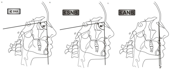
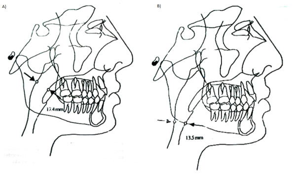
Materials and methods: This study was of the observational, descriptive, transversal, and prospective type. The sample was made up by 60 lateral head radiographs distributed between 30 class I (ANB 2°±2° and class I malocclusion) and 30 skeletal class II radiographs (ANB>5° and malocclusion class II-1). Measurements of the airway space in the oropharynx and nasopharynx were taken in mm through the McNamara method on lateral head radiographs.
Results: The average space found in the oropharynx in class I was 11.71mm ± 3.18mm. In the class II group, it was 10.73mm ± 2.36mm. No significant differences were found (p=0.18). The average space found in the nasopharynx in the class I group was 18.45mm ± 4.11mm. In the class II group, it was 19.10mm ± 3.89mm. There were no significant differences found (p=0.53).
Conclusion: The airway space in mm. of the nasopharynx presents similar values in millimeters in subjects with Class I and Class II skeletal malocclusion. There is no difference in the airway spaces of the oropharynx in subjects with Class I and Class II Malocclusions.

 求助内容:
求助内容: 应助结果提醒方式:


