Jordan M. Renna, Katelyn B. Sondereker, Christopher L. Cors, Sara N. Chaszeyka, Kristin N. Keenan, Michael R. Corigliano, Lindsey A. Milgrom, Jessica R. Onyak, Edward J. Hamad, Maureen E. Stabio
{"title":"From 2D slices to a 3D model: Training students in digital microanatomy analysis techniques through a 3D printed neuron project","authors":"Jordan M. Renna, Katelyn B. Sondereker, Christopher L. Cors, Sara N. Chaszeyka, Kristin N. Keenan, Michael R. Corigliano, Lindsey A. Milgrom, Jessica R. Onyak, Edward J. Hamad, Maureen E. Stabio","doi":"10.1002/ase.2396","DOIUrl":null,"url":null,"abstract":"<p>The reconstruction of two-dimensional (2D) slices to three-dimensional (3D) digital anatomical models requires technical skills and software that are becoming increasingly important to the modern anatomist, but these skills are rarely taught in undergraduate science classrooms. Furthermore, learning opportunities that allow students to simultaneously explore anatomy in both 2D and 3D space are increasingly valuable. This report describes a novel learning activity that trains students to digitally trace a serially imaged neuron from a confocal stack and to model that neuron in 3D space for 3D printing. By engaging students in the production of a 3D digital model, this learning activity is designed to provide students a novel way to enhance their understanding of the content, including didactic knowledge of neuron morphology, technical research skills in image analysis, and career exploration of neuroanatomy research. Moreover, students engage with microanatomy in a way that starts in 2D but results in a 3D object they can see, touch, and keep. This discursive article presents the learning activity, including videos, instructional guides, and learning objectives designed to engage students on all six levels of Bloom's Taxonomy. Furthermore, this work is a proof of principle modeling workflow that is approachable, inexpensive, achievable, and adaptable to cell types in other organ systems. This work is designed to motivate the expansion of 3D printing technology into microanatomy and neuroanatomy education.</p>","PeriodicalId":124,"journal":{"name":"Anatomical Sciences Education","volume":null,"pages":null},"PeriodicalIF":5.2000,"publicationDate":"2024-02-20","publicationTypes":"Journal Article","fieldsOfStudy":null,"isOpenAccess":false,"openAccessPdf":"https://onlinelibrary.wiley.com/doi/epdf/10.1002/ase.2396","citationCount":"0","resultStr":null,"platform":"Semanticscholar","paperid":null,"PeriodicalName":"Anatomical Sciences Education","FirstCategoryId":"95","ListUrlMain":"https://onlinelibrary.wiley.com/doi/10.1002/ase.2396","RegionNum":2,"RegionCategory":"教育学","ArticlePicture":[],"TitleCN":null,"AbstractTextCN":null,"PMCID":null,"EPubDate":"","PubModel":"","JCR":"Q1","JCRName":"EDUCATION, SCIENTIFIC DISCIPLINES","Score":null,"Total":0}
引用次数: 0
Abstract
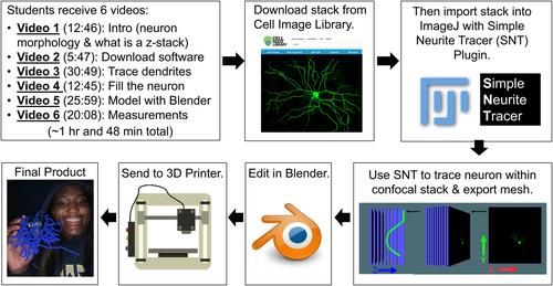
The reconstruction of two-dimensional (2D) slices to three-dimensional (3D) digital anatomical models requires technical skills and software that are becoming increasingly important to the modern anatomist, but these skills are rarely taught in undergraduate science classrooms. Furthermore, learning opportunities that allow students to simultaneously explore anatomy in both 2D and 3D space are increasingly valuable. This report describes a novel learning activity that trains students to digitally trace a serially imaged neuron from a confocal stack and to model that neuron in 3D space for 3D printing. By engaging students in the production of a 3D digital model, this learning activity is designed to provide students a novel way to enhance their understanding of the content, including didactic knowledge of neuron morphology, technical research skills in image analysis, and career exploration of neuroanatomy research. Moreover, students engage with microanatomy in a way that starts in 2D but results in a 3D object they can see, touch, and keep. This discursive article presents the learning activity, including videos, instructional guides, and learning objectives designed to engage students on all six levels of Bloom's Taxonomy. Furthermore, this work is a proof of principle modeling workflow that is approachable, inexpensive, achievable, and adaptable to cell types in other organ systems. This work is designed to motivate the expansion of 3D printing technology into microanatomy and neuroanatomy education.
将二维(2D)切片重建为三维(3D)数字解剖模型需要对现代解剖学家越来越重要的技术技能和软件,但这些技能很少在本科科学课堂上教授。此外,能让学生同时在二维和三维空间中探索解剖学的学习机会也越来越有价值。本报告介绍了一种新颖的学习活动,它训练学生从共聚焦堆栈中以数字方式追踪连续成像的神经元,并在三维空间中为该神经元建模,以便进行三维打印。通过让学生参与制作三维数字模型,该学习活动旨在为学生提供一种新颖的方式来加深对教学内容的理解,包括神经元形态学的教学知识、图像分析的技术研究技能以及神经解剖学研究的职业探索。此外,学生参与显微解剖学的方式始于二维,但结果却是他们可以看到、触摸和保存的三维物体。这篇论述性文章介绍了学习活动,包括视频、教学指南和学习目标,旨在让学生参与布卢姆分类学的所有六个层次。此外,这项工作还证明了建模工作流程的原理,该流程平易近人、成本低廉、易于实现,并可适用于其他器官系统的细胞类型。这项工作旨在推动 3D 打印技术在显微解剖学和神经解剖学教育中的应用。
期刊介绍:
Anatomical Sciences Education, affiliated with the American Association for Anatomy, serves as an international platform for sharing ideas, innovations, and research related to education in anatomical sciences. Covering gross anatomy, embryology, histology, and neurosciences, the journal addresses education at various levels, including undergraduate, graduate, post-graduate, allied health, medical (both allopathic and osteopathic), and dental. It fosters collaboration and discussion in the field of anatomical sciences education.
 求助内容:
求助内容: 应助结果提醒方式:


