Preoperative photoacoustic versus indocyanine green lymphography in lymphaticovenular anastomosis outcomes for lower extremity lymphedema: A pilot study
Abstract
Background
Identification of the proper lymphatics is important for successful lymphaticovenular anastomosis (LVA) for lymphedema; however, visualization of lymphatic vessels is challenging. Photoacoustic lymphangiography (PAL) can help visualize lymphatics more clearly than other modalities. Therefore, we investigated the usefulness of PAL and determined whether the clear and three-dimensional image of PAL affects LVA outcomes.
Methods
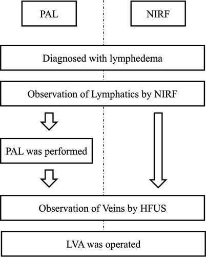
We recruited 22 female patients with lower extremity lymphedema. The operative time, number of incisions, number of anastomoses, lymphatic vessel detection rate (number of functional lymphatics identified during the operation/number of incisions), and limb volume changes preoperatively and 3 months postoperatively were compared retrospectively. The patients were divided according to whether PAL was performed or not, and results were compared between those undergoing PAL (PAL group; n = 10) and those who did not (near-infrared fluorescence [NIRF] group, n = 12).
Results
The mean age of the patients was 55.9 ± 15.1 years in the PAL group and 50.7 ± 14.9 years in the NIRF group. One patient in the PAL group and three in the NIRF group had primary lymphedema. Eighteen patients (PAL group, nine; and NIRF group, nine) had secondary lymphedema. Based on preoperative evaluation using the International Society of Lymphology (ISL) classification, eight patients were determined to be in stage 2 and two patients in late stage 2 in the PAL group. In contrast, in the NIRF group, one patient was determined to be in stage 0, three patients each in stage 1 and stage 2, and five patients in late stage 2.
Lymphatic vessel detection rates were 93% (42 LVAs and 45 incisions) and 83% (50 LVAs and 60 incisions) in the groups with and without PAL, respectively (p = 0.42). Limb volume change was evaluated in five limbs of four patients and in seven limbs of five patients in the PAL and NIRF groups as 336.6 ± 203.6 mL (5.90% ± 3.27%) and 52.9 ± 260.7 mL (0.71% ± 4.27%), respectively. The PAL group showed a significant volume reduction. (p = .038).
Conclusions
Detection of functional lymphatic vessels on PAL is useful for treating LVA.


 求助内容:
求助内容: 应助结果提醒方式:


