A Raman topography imaging method toward assisting surgical tumor resection

Alexander Czaja, Alice J. Jiang, Matt Zacchary Blanco, Olga E. Eremina, Cristina Zavaleta
{"title":"A Raman topography imaging method toward assisting surgical tumor resection","authors":"Alexander Czaja, Alice J. Jiang, Matt Zacchary Blanco, Olga E. Eremina, Cristina Zavaleta","doi":"10.1038/s44303-024-00006-6","DOIUrl":null,"url":null,"abstract":"Achieving complete tumor resection upon initial surgical intervention can lead to better patient outcomes by making adjuvant treatments more efficacious and reducing the strain of repeat surgeries. Complete tumor resection can be difficult to confirm intraoperatively. Methods like touch preparation (TP) have been inconsistent for detecting residual malignant cell populations, and fatty specimens like breast cancer lumpectomies are too fatty to process for rapid histology. We propose a novel workflow of immunostaining and topographic surface imaging of freshly excised tissue to ensure complete resection using highly sensitive and spectrally separable surface-enhanced Raman scattering nanoparticles (SERS NPs) as the targeted contrast agent. Biomarker-targeting SERS NPs are ideal contrast agents for this application because their sensitivity enables rapid detection, and their narrow bands enable extensive intra-pixel multiplexing. The adaptive focus capabilities of an advanced Raman instrument, combined with our rotational accessory device for exposing each surface of the stained specimen to the objective lens, enable topographic mapping of complete excised specimen surfaces. A USB-controlled accessory for a Raman microscope was designed and fabricated to enable programmatic and precise angular manipulation of specimens in concert with instrument stage motions during whole-surface imaging. Specimens are affixed to the accessory on an anti-slip, sterilizable rod, and the tissue surface exposed to the instrument is adjusted on demand using a programmed rotating stepper motor. We demonstrate this topographic imaging strategy on a variety of phantoms and preclinical tissue specimens. The results show detail and texture in specimen surface topography, orientation of findings and navigability across surfaces, and extensive SERS NP multiplexing and linear quantitation capabilities under this new Raman topography imaging method. We demonstrate successful surface mapping and recognition of all 26 of our distinct SERS NP types along with effective deconvolution and localization of randomly assigned NP mixtures. Increasing NP concentrations were also quantitatively assessed and showed a linear correlation with Raman signal with an R2 coefficient of determination of 0.97. Detailed surface renderings color-encoded by unmixed SERS NP abundances show a path forward for content-rich, interactive surgical margin assessment.","PeriodicalId":501709,"journal":{"name":"npj Imaging","volume":" ","pages":"1-11"},"PeriodicalIF":0.0000,"publicationDate":"2024-02-19","publicationTypes":"Journal Article","fieldsOfStudy":null,"isOpenAccess":false,"openAccessPdf":"https://www.nature.com/articles/s44303-024-00006-6.pdf","citationCount":"0","resultStr":null,"platform":"Semanticscholar","paperid":null,"PeriodicalName":"npj Imaging","FirstCategoryId":"1085","ListUrlMain":"https://www.nature.com/articles/s44303-024-00006-6","RegionNum":0,"RegionCategory":null,"ArticlePicture":[],"TitleCN":null,"AbstractTextCN":null,"PMCID":null,"EPubDate":"","PubModel":"","JCR":"","JCRName":"","Score":null,"Total":0}
引用次数: 0

Abstract

Achieving complete tumor resection upon initial surgical intervention can lead to better patient outcomes by making adjuvant treatments more efficacious and reducing the strain of repeat surgeries. Complete tumor resection can be difficult to confirm intraoperatively. Methods like touch preparation (TP) have been inconsistent for detecting residual malignant cell populations, and fatty specimens like breast cancer lumpectomies are too fatty to process for rapid histology. We propose a novel workflow of immunostaining and topographic surface imaging of freshly excised tissue to ensure complete resection using highly sensitive and spectrally separable surface-enhanced Raman scattering nanoparticles (SERS NPs) as the targeted contrast agent. Biomarker-targeting SERS NPs are ideal contrast agents for this application because their sensitivity enables rapid detection, and their narrow bands enable extensive intra-pixel multiplexing. The adaptive focus capabilities of an advanced Raman instrument, combined with our rotational accessory device for exposing each surface of the stained specimen to the objective lens, enable topographic mapping of complete excised specimen surfaces. A USB-controlled accessory for a Raman microscope was designed and fabricated to enable programmatic and precise angular manipulation of specimens in concert with instrument stage motions during whole-surface imaging. Specimens are affixed to the accessory on an anti-slip, sterilizable rod, and the tissue surface exposed to the instrument is adjusted on demand using a programmed rotating stepper motor. We demonstrate this topographic imaging strategy on a variety of phantoms and preclinical tissue specimens. The results show detail and texture in specimen surface topography, orientation of findings and navigability across surfaces, and extensive SERS NP multiplexing and linear quantitation capabilities under this new Raman topography imaging method. We demonstrate successful surface mapping and recognition of all 26 of our distinct SERS NP types along with effective deconvolution and localization of randomly assigned NP mixtures. Increasing NP concentrations were also quantitatively assessed and showed a linear correlation with Raman signal with an R2 coefficient of determination of 0.97. Detailed surface renderings color-encoded by unmixed SERS NP abundances show a path forward for content-rich, interactive surgical margin assessment.
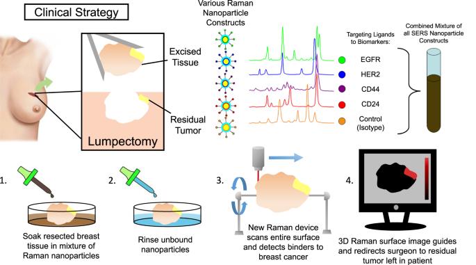
Abstract Image

用于辅助外科肿瘤切除的拉曼地形图成像方法
在初次手术干预时实现肿瘤的完全切除,可使辅助治疗更有效,并减少重复手术的压力,从而改善患者的预后。术中很难确认肿瘤是否完全切除。触摸制备(TP)等方法在检测残留恶性细胞群方面并不一致,而乳腺癌肿块切除术等脂肪标本过于肥厚,无法进行快速组织学处理。我们提出了一种对新鲜切除组织进行免疫染色和表面形貌成像的新工作流程,以确保使用高灵敏度和光谱可分离的表面增强拉曼散射纳米粒子(SERS NPs)作为靶向造影剂进行完整切除。生物标记物靶向 SERS NPs 是这一应用的理想造影剂,因为其灵敏度高,可实现快速检测,而且其窄波段可实现广泛的像素内复用。先进拉曼仪器的自适应聚焦功能与我们的旋转附件装置相结合,可将染色标本的每个表面都暴露在物镜下,从而对完整切除的标本表面进行地形图绘制。我们设计并制造了拉曼显微镜的 USB 控制附件,以便在全表面成像过程中配合仪器平台运动,对标本进行程序化的精确角度操作。标本被固定在一个防滑、可消毒的杆上,暴露在仪器上的组织表面可根据需要通过编程旋转步进电机进行调整。我们在各种模型和临床前组织标本上演示了这种地形成像策略。结果显示,在这种新型拉曼地形成像方法下,标本表面地形的细节和纹理、研究结果的方向和表面的可浏览性,以及广泛的 SERS NP 多路复用和线性定量能力。我们展示了所有 26 种不同 SERS NP 类型的成功表面映射和识别,以及随机分配的 NP 混合物的有效解卷积和定位。我们还对 NP 浓度的增加进行了定量评估,结果表明 NP 浓度与拉曼信号呈线性相关,R2 决定系数为 0.97。由未混合的 SERS NP 丰度编码的详细表面渲染为内容丰富的交互式手术边缘评估指明了方向。
本文章由计算机程序翻译,如有差异,请以英文原文为准。
求助全文
约1分钟内获得全文 求助全文
来源期刊
自引率
0.00%
发文量
0
×
引用
GB/T 7714-2015
复制
MLA
复制
APA
复制
导出至
BibTeX EndNote RefMan NoteFirst NoteExpress
×
提示
您的信息不完整,为了账户安全,请先补充。
现在去补充
×
提示
您因"违规操作"
具体请查看互助需知
我知道了
×
提示
确定
请完成安全验证×
copy
已复制链接
快去分享给好友吧!
我知道了
右上角分享
点击右上角分享
0
联系我们:info@booksci.cn Book学术提供免费学术资源搜索服务,方便国内外学者检索中英文文献。致力于提供最便捷和优质的服务体验。 Copyright © 2023 布克学术 All rights reserved.
京ICP备2023020795号-1
ghs 京公网安备 11010802042870号
Book学术文献互助
Book学术文献互助群
群 号:481959085
Book学术官方微信