Jianzhu Wei, Yang Zhang, Bo Xie, Ziyi Zhu, Jingyu Qian, Yulin Tan
{"title":"The atypical protein kinase RIOK3 contributes to the phenotypic modulation of vascular smooth muscle cells in intracranial aneurysms","authors":"Jianzhu Wei, Yang Zhang, Bo Xie, Ziyi Zhu, Jingyu Qian, Yulin Tan","doi":"10.1007/s13273-023-00425-3","DOIUrl":null,"url":null,"abstract":"<h3 data-test=\"abstract-sub-heading\">Background</h3><p>Previous studies manifested that abnormal proliferation, migration, apoptosis, and phenotypic conversion of vascular smooth muscle cells (VSMCs) are the main pathogenic basis of intracranial aneurysms (IAs).</p><h3 data-test=\"abstract-sub-heading\">Objective</h3><p>The aim of this study was to explore a key gene associated with IA growth and rupture using bioinformatics analysis and validate it by exogenous overexpression into human brain VSMCs (HBVSMCs). Four IA-associated microarray datasets, GSE54083, GSE15629, GSE66238, and GSE13353, were obtained from Gene Expression Omnibus (GEO) and analyzed using GEO2R for differentially expressed genes (DEGs). HBVSMCs were infected with lentivirus containing RIO kinase 3 (RIOK3) to overexpress exogenous RIOK3, and then, CCK-8, EdU, cell scratch, Transwell, Western blotting, and ELISA were introduced to measure proliferation, migration, phenotypic conversion-related proteins, and proinflammatory cytokines in HBVSMCs. To simulate the abnormal hemodynamic environment in the late stages of IA formation, RIOK3-overexpressing HBVSMCs were cultured under wall shear stress (WSS)-loaded conditions and then subjected to apoptosis assessment.</p><h3 data-test=\"abstract-sub-heading\">Results</h3><p>RIOK3 was defined as a key gene in the DEGs of IAs by bioinformatics analysis. RIOK3 overexpression could contribute to the abnormal proliferation, migration, secretion of proinflammatory factors, and the conversion of contractile phenotype to synthetic phenotype of HBVSMCs. Additionally, RIOK3 overexpression encouraged HBVSMC apoptosis after loading WSS in vitro to mimic advanced-IAs.</p><h3 data-test=\"abstract-sub-heading\">Conclusion</h3><p>RIOK3 in pre-IAs (without WSS loading) facilitates phenotypic conversion, abnormal proliferation, invasion, and inflammatory cytokine secretion of HBVSMCs; whereas in the advanced-IAs, RIOK3 accelerated the abnormal apoptosis of HBVSMCs in the setting of loaded-WSS.</p>","PeriodicalId":18683,"journal":{"name":"Molecular & Cellular Toxicology","volume":"18 1","pages":""},"PeriodicalIF":1.1000,"publicationDate":"2024-02-09","publicationTypes":"Journal Article","fieldsOfStudy":null,"isOpenAccess":false,"openAccessPdf":"","citationCount":"0","resultStr":null,"platform":"Semanticscholar","paperid":null,"PeriodicalName":"Molecular & Cellular Toxicology","FirstCategoryId":"3","ListUrlMain":"https://doi.org/10.1007/s13273-023-00425-3","RegionNum":4,"RegionCategory":"医学","ArticlePicture":[],"TitleCN":null,"AbstractTextCN":null,"PMCID":null,"EPubDate":"","PubModel":"","JCR":"Q4","JCRName":"TOXICOLOGY","Score":null,"Total":0}
引用次数: 0
Abstract
Background
Previous studies manifested that abnormal proliferation, migration, apoptosis, and phenotypic conversion of vascular smooth muscle cells (VSMCs) are the main pathogenic basis of intracranial aneurysms (IAs).
Objective
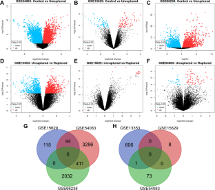
The aim of this study was to explore a key gene associated with IA growth and rupture using bioinformatics analysis and validate it by exogenous overexpression into human brain VSMCs (HBVSMCs). Four IA-associated microarray datasets, GSE54083, GSE15629, GSE66238, and GSE13353, were obtained from Gene Expression Omnibus (GEO) and analyzed using GEO2R for differentially expressed genes (DEGs). HBVSMCs were infected with lentivirus containing RIO kinase 3 (RIOK3) to overexpress exogenous RIOK3, and then, CCK-8, EdU, cell scratch, Transwell, Western blotting, and ELISA were introduced to measure proliferation, migration, phenotypic conversion-related proteins, and proinflammatory cytokines in HBVSMCs. To simulate the abnormal hemodynamic environment in the late stages of IA formation, RIOK3-overexpressing HBVSMCs were cultured under wall shear stress (WSS)-loaded conditions and then subjected to apoptosis assessment.
Results
RIOK3 was defined as a key gene in the DEGs of IAs by bioinformatics analysis. RIOK3 overexpression could contribute to the abnormal proliferation, migration, secretion of proinflammatory factors, and the conversion of contractile phenotype to synthetic phenotype of HBVSMCs. Additionally, RIOK3 overexpression encouraged HBVSMC apoptosis after loading WSS in vitro to mimic advanced-IAs.
Conclusion
RIOK3 in pre-IAs (without WSS loading) facilitates phenotypic conversion, abnormal proliferation, invasion, and inflammatory cytokine secretion of HBVSMCs; whereas in the advanced-IAs, RIOK3 accelerated the abnormal apoptosis of HBVSMCs in the setting of loaded-WSS.
期刊介绍:
Molecular & Cellular Toxicology publishes original research and reviews in all areas of the complex interaction between the cell´s genome (the sum of all genes within the chromosome), chemicals in the environment, and disease. Acceptable manuscripts are the ones that deal with some topics of environmental contaminants, including those that lie in the domains of analytical chemistry, biochemistry, pharmacology and toxicology with the aspects of molecular and cellular levels. Emphasis will be placed on toxic effects observed at relevant genomics and proteomics, which have direct impact on drug development, environment health, food safety, preventive medicine, and forensic medicine. The journal is committed to rapid peer review to ensure the publication of highest quality original research and timely news and review articles.
 求助内容:
求助内容: 应助结果提醒方式:


