Catherine Hatzantonis, Lalith Satkunam, Karyne N. Rabey, Jennifer C. Hocking, Anne M. R. Agur
{"title":"Fatty infiltration of gastrocnemius–soleus muscle complex: Considerations for myosteatosis rehabilitation","authors":"Catherine Hatzantonis, Lalith Satkunam, Karyne N. Rabey, Jennifer C. Hocking, Anne M. R. Agur","doi":"10.1111/joa.14025","DOIUrl":null,"url":null,"abstract":"<p>Although previous studies have reported fatty infiltration of the gastrocnemius–soleus complex, little is known about the volumetric distribution and patterns of fatty infiltration. The purpose of this anatomical study was to document and quantify the frequency, distribution, and pattern of fatty infiltration of the gastrocnemius–soleus complex. One hundred formalin-embalmed specimens (mean age 78.1 ± 12.3 years; 48F/52M) were serially dissected to document the frequency, distribution, and pattern of fatty infiltration in the medial and lateral heads of gastrocnemius and soleus muscles. Fatty infiltration was found in 23% of specimens, 13 unilaterally (8F/5M) and 10 (5M/5F) bilaterally. The fatty infiltration process was observed to begin medially from the medial aspect of the medial head of gastrocnemius and medial margin of soleus and then progressed laterally throughout the medial head of gastrocnemius and the marginal, anterior, and posterior soleus. The lateral head of gastrocnemius remained primarily muscular in all specimens. Microscopically, the pattern of infiltration was demonstrated as intramuscular with intact aponeuroses, and septa. The remaining endo-, peri-, and epimysium preserved the overall contour of the gastrocnemius–soleus complex, even in cases of significant fatty replacement. Since the external contour of the calf is preserved, the presence of fatty infiltration may be underdiagnosed in the clinic without imaging. Myosteatosis is associated with gait and balance challenges in the elderly, which can impact quality of life and result in increased risk of falling. The findings of the study have implications in the rehabilitation management of elderly patients with sarcopenia and myosteatosis.</p>","PeriodicalId":14971,"journal":{"name":"Journal of Anatomy","volume":null,"pages":null},"PeriodicalIF":1.8000,"publicationDate":"2024-02-16","publicationTypes":"Journal Article","fieldsOfStudy":null,"isOpenAccess":false,"openAccessPdf":"https://onlinelibrary.wiley.com/doi/epdf/10.1111/joa.14025","citationCount":"0","resultStr":null,"platform":"Semanticscholar","paperid":null,"PeriodicalName":"Journal of Anatomy","FirstCategoryId":"3","ListUrlMain":"https://onlinelibrary.wiley.com/doi/10.1111/joa.14025","RegionNum":3,"RegionCategory":"医学","ArticlePicture":[],"TitleCN":null,"AbstractTextCN":null,"PMCID":null,"EPubDate":"","PubModel":"","JCR":"Q2","JCRName":"ANATOMY & MORPHOLOGY","Score":null,"Total":0}
引用次数: 0
Abstract
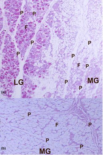
Although previous studies have reported fatty infiltration of the gastrocnemius–soleus complex, little is known about the volumetric distribution and patterns of fatty infiltration. The purpose of this anatomical study was to document and quantify the frequency, distribution, and pattern of fatty infiltration of the gastrocnemius–soleus complex. One hundred formalin-embalmed specimens (mean age 78.1 ± 12.3 years; 48F/52M) were serially dissected to document the frequency, distribution, and pattern of fatty infiltration in the medial and lateral heads of gastrocnemius and soleus muscles. Fatty infiltration was found in 23% of specimens, 13 unilaterally (8F/5M) and 10 (5M/5F) bilaterally. The fatty infiltration process was observed to begin medially from the medial aspect of the medial head of gastrocnemius and medial margin of soleus and then progressed laterally throughout the medial head of gastrocnemius and the marginal, anterior, and posterior soleus. The lateral head of gastrocnemius remained primarily muscular in all specimens. Microscopically, the pattern of infiltration was demonstrated as intramuscular with intact aponeuroses, and septa. The remaining endo-, peri-, and epimysium preserved the overall contour of the gastrocnemius–soleus complex, even in cases of significant fatty replacement. Since the external contour of the calf is preserved, the presence of fatty infiltration may be underdiagnosed in the clinic without imaging. Myosteatosis is associated with gait and balance challenges in the elderly, which can impact quality of life and result in increased risk of falling. The findings of the study have implications in the rehabilitation management of elderly patients with sarcopenia and myosteatosis.
期刊介绍:
Journal of Anatomy is an international peer-reviewed journal sponsored by the Anatomical Society. The journal publishes original papers, invited review articles and book reviews. Its main focus is to understand anatomy through an analysis of structure, function, development and evolution. Priority will be given to studies of that clearly articulate their relevance to the anatomical community. Focal areas include: experimental studies, contributions based on molecular and cell biology and on the application of modern imaging techniques and papers with novel methods or synthetic perspective on an anatomical system.
Studies that are essentially descriptive anatomy are appropriate only if they communicate clearly a broader functional or evolutionary significance. You must clearly state the broader implications of your work in the abstract.
We particularly welcome submissions in the following areas:
Cell biology and tissue architecture
Comparative functional morphology
Developmental biology
Evolutionary developmental biology
Evolutionary morphology
Functional human anatomy
Integrative vertebrate paleontology
Methodological innovations in anatomical research
Musculoskeletal system
Neuroanatomy and neurodegeneration
Significant advances in anatomical education.


 求助内容:
求助内容: 应助结果提醒方式:


