A method for crystallographic mapping of an alpha-beta titanium alloy with nanometre resolution using scanning precession electron diffraction and open-source software libraries
Ian MacLaren, Enrique Frutos-Myro, Steven Zeltmann, Colin Ophus
{"title":"A method for crystallographic mapping of an alpha-beta titanium alloy with nanometre resolution using scanning precession electron diffraction and open-source software libraries","authors":"Ian MacLaren, Enrique Frutos-Myro, Steven Zeltmann, Colin Ophus","doi":"10.1111/jmi.13275","DOIUrl":null,"url":null,"abstract":"<p>An approach for the crystallographic mapping of two-phase alloys on the nanoscale using a combination of scanned precession electron diffraction and open-source python libraries is introduced in this paper. This method is demonstrated using the example of a two-phase α/β titanium alloy. The data were recorded using a direct electron detector to collect the patterns, and recently developed algorithms to perform automated indexing and analyse the crystallography from the results. Very high-quality mapping is achieved at a 3 nm step size. The results show the expected Burgers orientation relationships between the α laths and β matrix, as well as the expected misorientations between α laths. A minor issue was found that one area was affected by 180° ambiguities in indexing occur due to this area being aligned too close to a zone axis of the α with twofold projection symmetry (not present in 3D) in the zero-order Laue Zone, and this should be avoided in data acquisition in the future. Nevertheless, this study demonstrates a good workflow for the analysis of nanocrystalline two- or multi-phase materials, which will be of widespread use in analysing two-phase titanium and other systems and how they evolve as a function of thermomechanical treatments.</p>","PeriodicalId":1,"journal":{"name":"Accounts of Chemical Research","volume":null,"pages":null},"PeriodicalIF":16.4000,"publicationDate":"2024-02-14","publicationTypes":"Journal Article","fieldsOfStudy":null,"isOpenAccess":false,"openAccessPdf":"https://onlinelibrary.wiley.com/doi/epdf/10.1111/jmi.13275","citationCount":"0","resultStr":null,"platform":"Semanticscholar","paperid":null,"PeriodicalName":"Accounts of Chemical Research","FirstCategoryId":"5","ListUrlMain":"https://onlinelibrary.wiley.com/doi/10.1111/jmi.13275","RegionNum":1,"RegionCategory":"化学","ArticlePicture":[],"TitleCN":null,"AbstractTextCN":null,"PMCID":null,"EPubDate":"","PubModel":"","JCR":"Q1","JCRName":"CHEMISTRY, MULTIDISCIPLINARY","Score":null,"Total":0}
引用次数: 0
Abstract
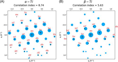
An approach for the crystallographic mapping of two-phase alloys on the nanoscale using a combination of scanned precession electron diffraction and open-source python libraries is introduced in this paper. This method is demonstrated using the example of a two-phase α/β titanium alloy. The data were recorded using a direct electron detector to collect the patterns, and recently developed algorithms to perform automated indexing and analyse the crystallography from the results. Very high-quality mapping is achieved at a 3 nm step size. The results show the expected Burgers orientation relationships between the α laths and β matrix, as well as the expected misorientations between α laths. A minor issue was found that one area was affected by 180° ambiguities in indexing occur due to this area being aligned too close to a zone axis of the α with twofold projection symmetry (not present in 3D) in the zero-order Laue Zone, and this should be avoided in data acquisition in the future. Nevertheless, this study demonstrates a good workflow for the analysis of nanocrystalline two- or multi-phase materials, which will be of widespread use in analysing two-phase titanium and other systems and how they evolve as a function of thermomechanical treatments.
期刊介绍:
Accounts of Chemical Research presents short, concise and critical articles offering easy-to-read overviews of basic research and applications in all areas of chemistry and biochemistry. These short reviews focus on research from the author’s own laboratory and are designed to teach the reader about a research project. In addition, Accounts of Chemical Research publishes commentaries that give an informed opinion on a current research problem. Special Issues online are devoted to a single topic of unusual activity and significance.
Accounts of Chemical Research replaces the traditional article abstract with an article "Conspectus." These entries synopsize the research affording the reader a closer look at the content and significance of an article. Through this provision of a more detailed description of the article contents, the Conspectus enhances the article's discoverability by search engines and the exposure for the research.

 求助内容:
求助内容: 应助结果提醒方式:


