Snehal Rajput , Rupal Kapdi , Mohendra Roy , Mehul S. Raval
{"title":"A triplanar ensemble model for brain tumor segmentation with volumetric multiparametric magnetic resonance images","authors":"Snehal Rajput , Rupal Kapdi , Mohendra Roy , Mehul S. Raval","doi":"10.1016/j.health.2024.100307","DOIUrl":null,"url":null,"abstract":"<div><p>Automated segmentation methods can produce faster segmentation of tumors in medical images, aiding medical professionals in diagnosis and treatment plans. A 3D U-Net method excels in this task but has high computational costs due to large model parameters, which limits their application under resource constraints. This study targets an optimized triplanar (2.5D) model ensemble to generate accurate segmentation with fewer parameters. The proposed triplanar model uses spatial and channel attention mechanisms and information from multiple orthogonal planar views to predict segmentation labels. In particular, we studied the optimum filter size to improve the accuracy without increasing the network complexity. The model generated output is further post-processed to fine-tune the segmentation results. The Dice similarity coefficients (Dice-score) of the Brain Tumor Segmentation (BraTS) 2020 training set for enhancing tumor (ET), whole tumor (WT), and tumor core (TC) are 0.736, 0.896, and 0.841, whereas, for the validation set, they are 0.713, 0.873, and 0.778, respectively. The proposed base model has only <span><math><mrow><mn>10</mn><mo>.</mo><mn>25</mn><mspace></mspace><mi>M</mi></mrow></math></span> parameters, three times less than BraTS 2020’s best-performing model (ET 0.798, WT 0.912, TC 0.857) on the validation set. The proposed ensemble model has <span><math><mrow><mn>93</mn><mo>.</mo><mn>5</mn><mspace></mspace><mi>M</mi></mrow></math></span> parameters, 1.6 times less than the top-ranked model and two times less than the third-ranked model (ET 0.793, WT 0.911, TC 0.853 on validation set) of BraTS2020 challenge.</p></div>","PeriodicalId":73222,"journal":{"name":"Healthcare analytics (New York, N.Y.)","volume":"5 ","pages":"Article 100307"},"PeriodicalIF":0.0000,"publicationDate":"2024-02-04","publicationTypes":"Journal Article","fieldsOfStudy":null,"isOpenAccess":false,"openAccessPdf":"https://www.sciencedirect.com/science/article/pii/S2772442524000091/pdfft?md5=d29fc0533e483abd517c7cab8004bdcb&pid=1-s2.0-S2772442524000091-main.pdf","citationCount":"0","resultStr":null,"platform":"Semanticscholar","paperid":null,"PeriodicalName":"Healthcare analytics (New York, N.Y.)","FirstCategoryId":"1085","ListUrlMain":"https://www.sciencedirect.com/science/article/pii/S2772442524000091","RegionNum":0,"RegionCategory":null,"ArticlePicture":[],"TitleCN":null,"AbstractTextCN":null,"PMCID":null,"EPubDate":"","PubModel":"","JCR":"","JCRName":"","Score":null,"Total":0}
引用次数: 0
Abstract
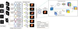
Automated segmentation methods can produce faster segmentation of tumors in medical images, aiding medical professionals in diagnosis and treatment plans. A 3D U-Net method excels in this task but has high computational costs due to large model parameters, which limits their application under resource constraints. This study targets an optimized triplanar (2.5D) model ensemble to generate accurate segmentation with fewer parameters. The proposed triplanar model uses spatial and channel attention mechanisms and information from multiple orthogonal planar views to predict segmentation labels. In particular, we studied the optimum filter size to improve the accuracy without increasing the network complexity. The model generated output is further post-processed to fine-tune the segmentation results. The Dice similarity coefficients (Dice-score) of the Brain Tumor Segmentation (BraTS) 2020 training set for enhancing tumor (ET), whole tumor (WT), and tumor core (TC) are 0.736, 0.896, and 0.841, whereas, for the validation set, they are 0.713, 0.873, and 0.778, respectively. The proposed base model has only parameters, three times less than BraTS 2020’s best-performing model (ET 0.798, WT 0.912, TC 0.857) on the validation set. The proposed ensemble model has parameters, 1.6 times less than the top-ranked model and two times less than the third-ranked model (ET 0.793, WT 0.911, TC 0.853 on validation set) of BraTS2020 challenge.

 求助内容:
求助内容: 应助结果提醒方式:


