P M Punithavathy, Ramesh Babu Telugu, Vinay Murahari Rao, Savit B Prabhu, Jayakanthan Kabeerdoss, Chanduni Syed, George Joseph, Debashish Danda, Meera Thomas, Ruchika Goel
{"title":"Study of pathogenic T-helper cell subsets in Asian Indian patients with Takayasu arteritis.","authors":"P M Punithavathy, Ramesh Babu Telugu, Vinay Murahari Rao, Savit B Prabhu, Jayakanthan Kabeerdoss, Chanduni Syed, George Joseph, Debashish Danda, Meera Thomas, Ruchika Goel","doi":"10.1007/s12026-024-09459-8","DOIUrl":null,"url":null,"abstract":"<p><p>The relapses and refractory disease are a challenge in the management of patients with Takayasu arteritis (TAK). We quantified pathogenic CD4 + memory T helper cells bearing surface markers CD161 and/or p-glycoprotein (MDR1) in patients with TAK. Peripheral blood mononuclear cells of 21 patients with TAK and 16 age-matched controls were stained with anti-CD3, anti-CD4, anti-CD45RA, anti-CD161 and anti-p-glycoprotein antibodies and subjected to flow cytometry by FACS ARIAIII. Eighteen patients underwent follow-up immunophenotyping. Intracellular staining for interleukin-17 and interferon-γ was performed for 18 patients and 11 controls. Surgical arterial biopsies of 6 TAK and 5 non-inflammatory controls were subjected to immunohistochemistry with anti-CD161 and anti-p-glycoprotein. At baseline the frequency of MDR1 + CD4 + and CD161 + MDR1 + CD4 + memory T cells was higher in TAK than controls (p = 0.002 and 0.01, respectively). After stimulation, the frequency of IFN-y + CD161 + cells was higher in TAK than controls (p = 0.028). Modal fluorescence intensity of CD161 + MDR1 + CD45RA - CD4 + cells was higher in active as compared with stable disease (p = 0.041). At 6 months, MDR1 + and CD161 + MDR1 + memory CD4 + T cells decreased significantly only in patients who had complete/partial response to treatment (p = 0.047 and 0.02, respectively). To conclude, MDR1 + and MDR1 + CD161 + CD4 + memory T-helper cells are increased in patients with TAK. These cells decreased only in patients with response to treatment during subsequent follow-up.</p>","PeriodicalId":13389,"journal":{"name":"Immunologic Research","volume":" ","pages":"636-643"},"PeriodicalIF":3.1000,"publicationDate":"2024-08-01","publicationTypes":"Journal Article","fieldsOfStudy":null,"isOpenAccess":false,"openAccessPdf":"","citationCount":"0","resultStr":null,"platform":"Semanticscholar","paperid":null,"PeriodicalName":"Immunologic Research","FirstCategoryId":"3","ListUrlMain":"https://doi.org/10.1007/s12026-024-09459-8","RegionNum":4,"RegionCategory":"医学","ArticlePicture":[],"TitleCN":null,"AbstractTextCN":null,"PMCID":null,"EPubDate":"2024/2/8 0:00:00","PubModel":"Epub","JCR":"Q3","JCRName":"IMMUNOLOGY","Score":null,"Total":0}
引用次数: 0
Abstract
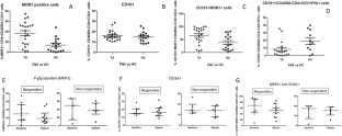
The relapses and refractory disease are a challenge in the management of patients with Takayasu arteritis (TAK). We quantified pathogenic CD4 + memory T helper cells bearing surface markers CD161 and/or p-glycoprotein (MDR1) in patients with TAK. Peripheral blood mononuclear cells of 21 patients with TAK and 16 age-matched controls were stained with anti-CD3, anti-CD4, anti-CD45RA, anti-CD161 and anti-p-glycoprotein antibodies and subjected to flow cytometry by FACS ARIAIII. Eighteen patients underwent follow-up immunophenotyping. Intracellular staining for interleukin-17 and interferon-γ was performed for 18 patients and 11 controls. Surgical arterial biopsies of 6 TAK and 5 non-inflammatory controls were subjected to immunohistochemistry with anti-CD161 and anti-p-glycoprotein. At baseline the frequency of MDR1 + CD4 + and CD161 + MDR1 + CD4 + memory T cells was higher in TAK than controls (p = 0.002 and 0.01, respectively). After stimulation, the frequency of IFN-y + CD161 + cells was higher in TAK than controls (p = 0.028). Modal fluorescence intensity of CD161 + MDR1 + CD45RA - CD4 + cells was higher in active as compared with stable disease (p = 0.041). At 6 months, MDR1 + and CD161 + MDR1 + memory CD4 + T cells decreased significantly only in patients who had complete/partial response to treatment (p = 0.047 and 0.02, respectively). To conclude, MDR1 + and MDR1 + CD161 + CD4 + memory T-helper cells are increased in patients with TAK. These cells decreased only in patients with response to treatment during subsequent follow-up.
期刊介绍:
IMMUNOLOGIC RESEARCH represents a unique medium for the presentation, interpretation, and clarification of complex scientific data. Information is presented in the form of interpretive synthesis reviews, original research articles, symposia, editorials, and theoretical essays. The scope of coverage extends to cellular immunology, immunogenetics, molecular and structural immunology, immunoregulation and autoimmunity, immunopathology, tumor immunology, host defense and microbial immunity, including viral immunology, immunohematology, mucosal immunity, complement, transplantation immunology, clinical immunology, neuroimmunology, immunoendocrinology, immunotoxicology, translational immunology, and history of immunology.
 求助内容:
求助内容: 应助结果提醒方式:


