JoonNyung Heo, Yongsik Sim, Byung Moon Kim, Dong Joon Kim, Young Dae Kim, Hyo Suk Nam, Yoon Seong Choi, Seung-Koo Lee, Eung Yeop Kim, Beomseok Sohn
{"title":"Radiomics using non-contrast CT to predict hemorrhagic transformation risk in stroke patients undergoing revascularization","authors":"JoonNyung Heo, Yongsik Sim, Byung Moon Kim, Dong Joon Kim, Young Dae Kim, Hyo Suk Nam, Yoon Seong Choi, Seung-Koo Lee, Eung Yeop Kim, Beomseok Sohn","doi":"10.1007/s00330-024-10618-6","DOIUrl":null,"url":null,"abstract":"<h3 data-test=\"abstract-sub-heading\">Objectives</h3><p>This study explores whether textural features from initial non-contrast CT scans of infarcted brain tissue are linked to hemorrhagic transformation susceptibility.</p><h3 data-test=\"abstract-sub-heading\">Materials and methods</h3><p>Stroke patients undergoing thrombolysis or thrombectomy from Jan 2012 to Jan 2022 were analyzed retrospectively. Hemorrhagic transformation was defined using follow-up magnetic resonance imaging. A total of 94 radiomic features were extracted from the infarcted tissue on initial NCCT scans. Patients were divided into training and test sets (7:3 ratio). Two models were developed with fivefold cross-validation: one incorporating first-order and textural radiomic features, and another using only textural radiomic features. A clinical model was also constructed using logistic regression with clinical variables, and test set validation was performed.</p><h3 data-test=\"abstract-sub-heading\">Results</h3><p>Among 362 patients, 218 had hemorrhagic transformations. The LightGBM model with all radiomics features had the best performance, with an area under the receiver operating characteristic curve (AUROC) of 0.986 (95% confidence interval [CI], 0.971–1.000) on the test dataset. The ExtraTrees model performed best when textural features were employed, with an AUROC of 0.845 (95% CI, 0.774–0.916). Minimum, maximum, and ten percentile values were significant predictors of hemorrhagic transformation. The clinical model showed an AUROC of 0.544 (95% CI, 0.431–0.658). The performance of the radiomics models was significantly better than that of the clinical model on the test dataset (<i>p</i> < 0.001).</p><h3 data-test=\"abstract-sub-heading\">Conclusions</h3><p>The radiomics model can predict hemorrhagic transformation using NCCT in stroke patients. Low Hounsfield unit was a strong predictor of hemorrhagic transformation, while textural features alone can predict hemorrhagic transformation.</p><h3 data-test=\"abstract-sub-heading\">Clinical relevance statement</h3><p>Using radiomic features extracted from initial non-contrast computed tomography, early prediction of hemorrhagic transformation has the potential to improve patient care and outcomes by aiding in personalized treatment decision-making and early identification of at-risk patients.</p><h3 data-test=\"abstract-sub-heading\">Key Points</h3><p><i>• Predicting hemorrhagic transformation following thrombolysis in stroke is challenging since multiple factors are associated.</i></p><p><i>• Radiomics features of infarcted tissue on initial non-contrast CT are associated with hemorrhagic transformation.</i></p><p><i>• Textural features on non-contrast CT are associated with the frailty of the infarcted tissue.</i></p>","PeriodicalId":12076,"journal":{"name":"European Radiology","volume":"10 1","pages":""},"PeriodicalIF":4.7000,"publicationDate":"2024-02-03","publicationTypes":"Journal Article","fieldsOfStudy":null,"isOpenAccess":false,"openAccessPdf":"","citationCount":"0","resultStr":null,"platform":"Semanticscholar","paperid":null,"PeriodicalName":"European Radiology","FirstCategoryId":"3","ListUrlMain":"https://doi.org/10.1007/s00330-024-10618-6","RegionNum":2,"RegionCategory":"医学","ArticlePicture":[],"TitleCN":null,"AbstractTextCN":null,"PMCID":null,"EPubDate":"","PubModel":"","JCR":"Q1","JCRName":"RADIOLOGY, NUCLEAR MEDICINE & MEDICAL IMAGING","Score":null,"Total":0}
引用次数: 0
Abstract
Objectives
This study explores whether textural features from initial non-contrast CT scans of infarcted brain tissue are linked to hemorrhagic transformation susceptibility.
Materials and methods
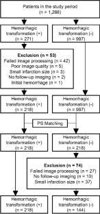
Stroke patients undergoing thrombolysis or thrombectomy from Jan 2012 to Jan 2022 were analyzed retrospectively. Hemorrhagic transformation was defined using follow-up magnetic resonance imaging. A total of 94 radiomic features were extracted from the infarcted tissue on initial NCCT scans. Patients were divided into training and test sets (7:3 ratio). Two models were developed with fivefold cross-validation: one incorporating first-order and textural radiomic features, and another using only textural radiomic features. A clinical model was also constructed using logistic regression with clinical variables, and test set validation was performed.
Results
Among 362 patients, 218 had hemorrhagic transformations. The LightGBM model with all radiomics features had the best performance, with an area under the receiver operating characteristic curve (AUROC) of 0.986 (95% confidence interval [CI], 0.971–1.000) on the test dataset. The ExtraTrees model performed best when textural features were employed, with an AUROC of 0.845 (95% CI, 0.774–0.916). Minimum, maximum, and ten percentile values were significant predictors of hemorrhagic transformation. The clinical model showed an AUROC of 0.544 (95% CI, 0.431–0.658). The performance of the radiomics models was significantly better than that of the clinical model on the test dataset (p < 0.001).
Conclusions
The radiomics model can predict hemorrhagic transformation using NCCT in stroke patients. Low Hounsfield unit was a strong predictor of hemorrhagic transformation, while textural features alone can predict hemorrhagic transformation.
Clinical relevance statement
Using radiomic features extracted from initial non-contrast computed tomography, early prediction of hemorrhagic transformation has the potential to improve patient care and outcomes by aiding in personalized treatment decision-making and early identification of at-risk patients.
Key Points
• Predicting hemorrhagic transformation following thrombolysis in stroke is challenging since multiple factors are associated.
• Radiomics features of infarcted tissue on initial non-contrast CT are associated with hemorrhagic transformation.
• Textural features on non-contrast CT are associated with the frailty of the infarcted tissue.
期刊介绍:
European Radiology (ER) continuously updates scientific knowledge in radiology by publication of strong original articles and state-of-the-art reviews written by leading radiologists. A well balanced combination of review articles, original papers, short communications from European radiological congresses and information on society matters makes ER an indispensable source for current information in this field.
This is the Journal of the European Society of Radiology, and the official journal of a number of societies.
From 2004-2008 supplements to European Radiology were published under its companion, European Radiology Supplements, ISSN 1613-3749.

 求助内容:
求助内容: 应助结果提醒方式:


