Megan Clapperton, Tash Kunanandam, Catalina D. Florea, Catriona M. Douglas, Gail McConnell
{"title":"Multimodal optical mesoscopy reveals the quantity and spatial distribution of Gram-positive biofilms in ex vivo tonsils","authors":"Megan Clapperton, Tash Kunanandam, Catalina D. Florea, Catriona M. Douglas, Gail McConnell","doi":"10.1111/jmi.13266","DOIUrl":null,"url":null,"abstract":"<p>Biofilms are known to be present in tonsils, but little is known about their spatial location and size distribution throughout the tonsil. Studies of the location and distribution of biofilms in tonsil specimens have thus far been limited to either high-magnification methods such as electron microscopy, which enables high-resolution imaging but only from a tiny tissue volume, or lower magnification techniques such as light microscopy, which allow imaging of larger specimens but with poor spatial resolution. To overcome these limitations, we report the use of multimodal optical mesoscopy to visualise and quantify the number and spatial distribution of Gram-positive biofilms in fresh, excised paediatric tonsils. This methodology supports simultaneous imaging of both the tonsil host and biofilms in whole mounts of tissue up to 5 mm × 5 mm × 3 mm with subcellular resolution throughout. A quantitative assessment of 36 tonsil specimens revealed no statistically significant difference between biofilm presence on the tonsil surface and the interior of the tonsil. This new quantitative mesoscale imaging approach may prove useful in understanding the role of biofilms in tonsillar diseases and other infections.</p>","PeriodicalId":1,"journal":{"name":"Accounts of Chemical Research","volume":null,"pages":null},"PeriodicalIF":16.4000,"publicationDate":"2024-01-31","publicationTypes":"Journal Article","fieldsOfStudy":null,"isOpenAccess":false,"openAccessPdf":"https://onlinelibrary.wiley.com/doi/epdf/10.1111/jmi.13266","citationCount":"0","resultStr":null,"platform":"Semanticscholar","paperid":null,"PeriodicalName":"Accounts of Chemical Research","FirstCategoryId":"5","ListUrlMain":"https://onlinelibrary.wiley.com/doi/10.1111/jmi.13266","RegionNum":1,"RegionCategory":"化学","ArticlePicture":[],"TitleCN":null,"AbstractTextCN":null,"PMCID":null,"EPubDate":"","PubModel":"","JCR":"Q1","JCRName":"CHEMISTRY, MULTIDISCIPLINARY","Score":null,"Total":0}
引用次数: 0
Abstract
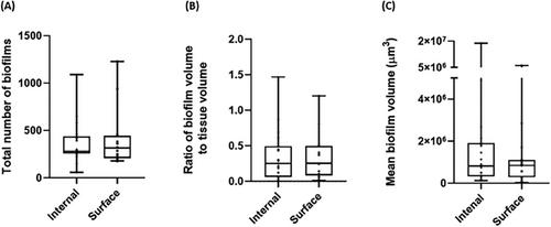
Biofilms are known to be present in tonsils, but little is known about their spatial location and size distribution throughout the tonsil. Studies of the location and distribution of biofilms in tonsil specimens have thus far been limited to either high-magnification methods such as electron microscopy, which enables high-resolution imaging but only from a tiny tissue volume, or lower magnification techniques such as light microscopy, which allow imaging of larger specimens but with poor spatial resolution. To overcome these limitations, we report the use of multimodal optical mesoscopy to visualise and quantify the number and spatial distribution of Gram-positive biofilms in fresh, excised paediatric tonsils. This methodology supports simultaneous imaging of both the tonsil host and biofilms in whole mounts of tissue up to 5 mm × 5 mm × 3 mm with subcellular resolution throughout. A quantitative assessment of 36 tonsil specimens revealed no statistically significant difference between biofilm presence on the tonsil surface and the interior of the tonsil. This new quantitative mesoscale imaging approach may prove useful in understanding the role of biofilms in tonsillar diseases and other infections.
期刊介绍:
Accounts of Chemical Research presents short, concise and critical articles offering easy-to-read overviews of basic research and applications in all areas of chemistry and biochemistry. These short reviews focus on research from the author’s own laboratory and are designed to teach the reader about a research project. In addition, Accounts of Chemical Research publishes commentaries that give an informed opinion on a current research problem. Special Issues online are devoted to a single topic of unusual activity and significance.
Accounts of Chemical Research replaces the traditional article abstract with an article "Conspectus." These entries synopsize the research affording the reader a closer look at the content and significance of an article. Through this provision of a more detailed description of the article contents, the Conspectus enhances the article's discoverability by search engines and the exposure for the research.

 求助内容:
求助内容: 应助结果提醒方式:


