{"title":"An automated slide scanning system for membrane filter imaging in diagnosis of urogenital schistosomiasis","authors":"Prosper Oyibo, Tope Agbana, Lisette van Lieshout, Wellington Oyibo, Jan-Carel Diehl, Gleb Vdovine","doi":"10.1111/jmi.13269","DOIUrl":null,"url":null,"abstract":"<p>Traditionally, automated slide scanning involves capturing a rectangular grid of field-of-view (FoV) images which can be stitched together to create whole slide images, while the autofocusing algorithm captures a focal stack of images to determine the best in-focus image. However, these methods can be time-consuming due to the need for <i>X</i>-, <i>Y</i>- and <i>Z</i>-axis movements of the digital microscope while capturing multiple FoV images. In this paper, we propose a solution to minimise these redundancies by presenting an optimal procedure for automated slide scanning of circular membrane filters on a glass slide. We achieve this by following an optimal path in the sample plane, ensuring that only FoVs overlapping the filter membrane are captured. To capture the best in-focus FoV image, we utilise a hill-climbing approach that tracks the peak of the mean of Gaussian gradient of the captured FoVs images along the <i>Z</i>-axis. We implemented this procedure to optimise the efficiency of the Schistoscope, an automated digital microscope developed to diagnose urogenital schistosomiasis by imaging <i>Schistosoma haematobium</i> eggs on 13 or 25 mm membrane filters. Our improved method reduces the automated slide scanning time by 63.18% and 72.52% for the respective filter sizes. This advancement greatly supports the practicality of the Schistoscope in large-scale schistosomiasis monitoring and evaluation programs in endemic regions. This will save time, resources and also accelerate generation of data that is critical in achieving the targets for schistosomiasis elimination.</p>","PeriodicalId":16484,"journal":{"name":"Journal of microscopy","volume":"294 1","pages":"52-61"},"PeriodicalIF":1.5000,"publicationDate":"2024-01-30","publicationTypes":"Journal Article","fieldsOfStudy":null,"isOpenAccess":false,"openAccessPdf":"https://onlinelibrary.wiley.com/doi/epdf/10.1111/jmi.13269","citationCount":"0","resultStr":null,"platform":"Semanticscholar","paperid":null,"PeriodicalName":"Journal of microscopy","FirstCategoryId":"5","ListUrlMain":"https://onlinelibrary.wiley.com/doi/10.1111/jmi.13269","RegionNum":4,"RegionCategory":"工程技术","ArticlePicture":[],"TitleCN":null,"AbstractTextCN":null,"PMCID":null,"EPubDate":"","PubModel":"","JCR":"Q3","JCRName":"MICROSCOPY","Score":null,"Total":0}
引用次数: 0
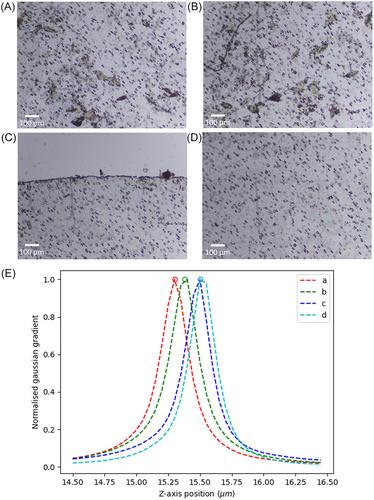
Abstract
Traditionally, automated slide scanning involves capturing a rectangular grid of field-of-view (FoV) images which can be stitched together to create whole slide images, while the autofocusing algorithm captures a focal stack of images to determine the best in-focus image. However, these methods can be time-consuming due to the need for X-, Y- and Z-axis movements of the digital microscope while capturing multiple FoV images. In this paper, we propose a solution to minimise these redundancies by presenting an optimal procedure for automated slide scanning of circular membrane filters on a glass slide. We achieve this by following an optimal path in the sample plane, ensuring that only FoVs overlapping the filter membrane are captured. To capture the best in-focus FoV image, we utilise a hill-climbing approach that tracks the peak of the mean of Gaussian gradient of the captured FoVs images along the Z-axis. We implemented this procedure to optimise the efficiency of the Schistoscope, an automated digital microscope developed to diagnose urogenital schistosomiasis by imaging Schistosoma haematobium eggs on 13 or 25 mm membrane filters. Our improved method reduces the automated slide scanning time by 63.18% and 72.52% for the respective filter sizes. This advancement greatly supports the practicality of the Schistoscope in large-scale schistosomiasis monitoring and evaluation programs in endemic regions. This will save time, resources and also accelerate generation of data that is critical in achieving the targets for schistosomiasis elimination.
期刊介绍:
The Journal of Microscopy is the oldest journal dedicated to the science of microscopy and the only peer-reviewed publication of the Royal Microscopical Society. It publishes papers that report on the very latest developments in microscopy such as advances in microscopy techniques or novel areas of application. The Journal does not seek to publish routine applications of microscopy or specimen preparation even though the submission may otherwise have a high scientific merit.
The scope covers research in the physical and biological sciences and covers imaging methods using light, electrons, X-rays and other radiations as well as atomic force and near field techniques. Interdisciplinary research is welcome. Papers pertaining to microscopy are also welcomed on optical theory, spectroscopy, novel specimen preparation and manipulation methods and image recording, processing and analysis including dynamic analysis of living specimens.
Publication types include full papers, hot topic fast tracked communications and review articles. Authors considering submitting a review article should contact the editorial office first.

 求助内容:
求助内容: 应助结果提醒方式:


