Zahra Setayesh-Mehr, Mohammad Hajitabar, Asghar Parsaei
{"title":"The Role of the HL-7 Peptide in the Induction of the Intrinsic Signalling Pathway of Apoptosis in HeLa Cancer Cells","authors":"Zahra Setayesh-Mehr, Mohammad Hajitabar, Asghar Parsaei","doi":"10.1134/S1990747823070036","DOIUrl":null,"url":null,"abstract":"<p>Anticancer peptides are of interest for cancer treatment. Nowadays, the process of apoptosis is considered a molecular target for cancer therapy. In the present study, the toxic effect of the HL-7 peptide on cervical cancer cells HeLa was investigated using the MTT assay. Also, the expression levels of <i>Bax</i>, <i>Bcl-2</i>, <i>p53</i>, <i>caspase-3</i>, <i>PTEN</i>, and <i>Akt</i> genes in HeLa cells treated with HL-7 were assessed by real-time PCR. Besides, the percentage of cells in early and late stages of apoptosis was determined using flow cytometry. The obtained results indicated that the peptide HL-7 inhibited growth of HeLa cells with IC<sub>50</sub> of 31 μM. The expression levels of <i>Bax</i>, <i>p53</i>, <i>caspase-3</i>, and <i>PTEN</i> genes were increased in HeLa cells treated with the HL-7 peptide as compared to untreated HeLa cells, while the expression levels of <i>Bcl-2</i> and <i>Akt</i> genes was decreased (<i>p</i> < 0.05). The results of flow cytometry analysis indicated a high percentage of cells in the late apoptosis stage (<i>p</i> < 0.05). Our findings suggest that peptide HL-7 can be involved in inducing the mitochondria-dependent apoptosis pathway. However, additional studies are needed to elucidate the exact mechanism of action of the peptide on HeLa cancer cells and the prospects for its therapeutic use in the clinic.</p>","PeriodicalId":484,"journal":{"name":"Biochemistry (Moscow), Supplement Series A: Membrane and Cell Biology","volume":"17 1 supplement","pages":"S78 - S84"},"PeriodicalIF":1.4000,"publicationDate":"2024-01-17","publicationTypes":"Journal Article","fieldsOfStudy":null,"isOpenAccess":false,"openAccessPdf":"","citationCount":"0","resultStr":null,"platform":"Semanticscholar","paperid":null,"PeriodicalName":"Biochemistry (Moscow), Supplement Series A: Membrane and Cell Biology","FirstCategoryId":"2","ListUrlMain":"https://link.springer.com/article/10.1134/S1990747823070036","RegionNum":0,"RegionCategory":null,"ArticlePicture":[],"TitleCN":null,"AbstractTextCN":null,"PMCID":null,"EPubDate":"","PubModel":"","JCR":"Q4","JCRName":"CELL BIOLOGY","Score":null,"Total":0}
引用次数: 0
Abstract
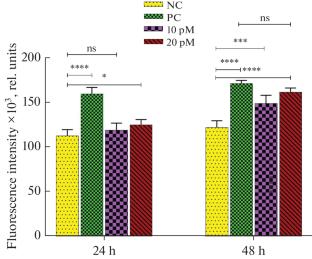
Anticancer peptides are of interest for cancer treatment. Nowadays, the process of apoptosis is considered a molecular target for cancer therapy. In the present study, the toxic effect of the HL-7 peptide on cervical cancer cells HeLa was investigated using the MTT assay. Also, the expression levels of Bax, Bcl-2, p53, caspase-3, PTEN, and Akt genes in HeLa cells treated with HL-7 were assessed by real-time PCR. Besides, the percentage of cells in early and late stages of apoptosis was determined using flow cytometry. The obtained results indicated that the peptide HL-7 inhibited growth of HeLa cells with IC50 of 31 μM. The expression levels of Bax, p53, caspase-3, and PTEN genes were increased in HeLa cells treated with the HL-7 peptide as compared to untreated HeLa cells, while the expression levels of Bcl-2 and Akt genes was decreased (p < 0.05). The results of flow cytometry analysis indicated a high percentage of cells in the late apoptosis stage (p < 0.05). Our findings suggest that peptide HL-7 can be involved in inducing the mitochondria-dependent apoptosis pathway. However, additional studies are needed to elucidate the exact mechanism of action of the peptide on HeLa cancer cells and the prospects for its therapeutic use in the clinic.
期刊介绍:
Biochemistry (Moscow), Supplement Series A: Membrane and Cell Biology is an international peer reviewed journal that publishes original articles on physical, chemical, and molecular mechanisms that underlie basic properties of biological membranes and mediate membrane-related cellular functions. The primary topics of the journal are membrane structure, mechanisms of membrane transport, bioenergetics and photobiology, intracellular signaling as well as membrane aspects of cell biology, immunology, and medicine. The journal is multidisciplinary and gives preference to those articles that employ a variety of experimental approaches, basically in biophysics but also in biochemistry, cytology, and molecular biology. The journal publishes articles that strive for unveiling membrane and cellular functions through innovative theoretical models and computer simulations.
 求助内容:
求助内容: 应助结果提醒方式:


