Cytologic features of canine melanotroph and corticotroph pituitary adenomas

IF 1.2 4区 农林科学 Q3 VETERINARY SCIENCES
Adriana P. Furtado, Michelle DeCourcey, Margaret A. Miller, Annie V. Chen, Linda G. Martin, Sarah C. Guess, K. Jane Wardrop, Cleverson de Souza, Tina J. Owen
{"title":"Cytologic features of canine melanotroph and corticotroph pituitary adenomas","authors":"Adriana P. Furtado,&nbsp;Michelle DeCourcey,&nbsp;Margaret A. Miller,&nbsp;Annie V. Chen,&nbsp;Linda G. Martin,&nbsp;Sarah C. Guess,&nbsp;K. Jane Wardrop,&nbsp;Cleverson de Souza,&nbsp;Tina J. Owen","doi":"10.1111/vcp.13311","DOIUrl":null,"url":null,"abstract":"<div>\n \n \n <section>\n \n <h3> Background</h3>\n \n <p>The introduction of intraoperative cytology revolutionized neurosurgical procedures in human medicine, providing real-time diagnostic guidance to surgeons and contributing to improved patient outcomes. In the realm of veterinary medicine, the understanding of pituitary tumors in dogs and cats remains limited due to challenges in obtaining antemortem samples of central nervous system lesions.</p>\n </section>\n \n <section>\n \n <h3> Objectives</h3>\n \n <p>The aim of this study was to describe the cytologic features of pituitary adenomas in 12 dogs that underwent hypophysectomy.</p>\n </section>\n \n <section>\n \n <h3> Methods</h3>\n \n <p>The series included nine melanotroph adenomas and three corticotroph adenomas. Definitive diagnosis was based on histopathology and immunohistochemistry.</p>\n </section>\n \n <section>\n \n <h3> Results</h3>\n \n <p>Cytologically, the adenomas had high numbers of bare nuclei and intact cells that were round to polygonal and situated individually or in small clusters. The intact cells had round to oval, eccentric nuclei with finely stippled chromatin and one to three prominent nucleoli and ample to abundant lightly basophilic to amphophilic, grainy cytoplasm with distinct borders, and variable numbers of discrete vacuoles. Mild-to-moderate anisocytosis and anisokaryosis, occasional binucleation, rare and atypical mitotic figures, and nuclear molding were also observed.</p>\n </section>\n \n <section>\n \n <h3> Conclusions</h3>\n \n <p>The results suggest that intraoperative cytology of canine pituitary adenomas holds promise as a valuable diagnostic tool, aiding swift differentiation from other sellar masses before histologic confirmation. Cytologic characterization of pituitary adenomas in dogs is exceptionally rare in the scientific literature, making this study one of the first to offer a comprehensive description of these cytologic features.</p>\n </section>\n </div>","PeriodicalId":23593,"journal":{"name":"Veterinary clinical pathology","volume":null,"pages":null},"PeriodicalIF":1.2000,"publicationDate":"2024-01-18","publicationTypes":"Journal Article","fieldsOfStudy":null,"isOpenAccess":false,"openAccessPdf":"https://onlinelibrary.wiley.com/doi/epdf/10.1111/vcp.13311","citationCount":"0","resultStr":null,"platform":"Semanticscholar","paperid":null,"PeriodicalName":"Veterinary clinical pathology","FirstCategoryId":"97","ListUrlMain":"https://onlinelibrary.wiley.com/doi/10.1111/vcp.13311","RegionNum":4,"RegionCategory":"农林科学","ArticlePicture":[],"TitleCN":null,"AbstractTextCN":null,"PMCID":null,"EPubDate":"","PubModel":"","JCR":"Q3","JCRName":"VETERINARY SCIENCES","Score":null,"Total":0}
引用次数: 0

Abstract

Background

The introduction of intraoperative cytology revolutionized neurosurgical procedures in human medicine, providing real-time diagnostic guidance to surgeons and contributing to improved patient outcomes. In the realm of veterinary medicine, the understanding of pituitary tumors in dogs and cats remains limited due to challenges in obtaining antemortem samples of central nervous system lesions.

Objectives

The aim of this study was to describe the cytologic features of pituitary adenomas in 12 dogs that underwent hypophysectomy.

Methods

The series included nine melanotroph adenomas and three corticotroph adenomas. Definitive diagnosis was based on histopathology and immunohistochemistry.

Results

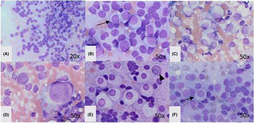
Cytologically, the adenomas had high numbers of bare nuclei and intact cells that were round to polygonal and situated individually or in small clusters. The intact cells had round to oval, eccentric nuclei with finely stippled chromatin and one to three prominent nucleoli and ample to abundant lightly basophilic to amphophilic, grainy cytoplasm with distinct borders, and variable numbers of discrete vacuoles. Mild-to-moderate anisocytosis and anisokaryosis, occasional binucleation, rare and atypical mitotic figures, and nuclear molding were also observed.

Conclusions

The results suggest that intraoperative cytology of canine pituitary adenomas holds promise as a valuable diagnostic tool, aiding swift differentiation from other sellar masses before histologic confirmation. Cytologic characterization of pituitary adenomas in dogs is exceptionally rare in the scientific literature, making this study one of the first to offer a comprehensive description of these cytologic features.
Abstract Image

犬黑质垂体腺瘤和皮质垂体腺瘤的细胞学特征。
背景:术中细胞学的引入彻底改变了人类医学中的神经外科手术,为外科医生提供了实时诊断指导,有助于改善患者预后。在兽医领域,由于难以获得中枢神经系统病变的死前样本,人们对犬猫垂体瘤的了解仍然有限:本研究旨在描述 12 只接受垂体下叶切除术的犬的垂体腺瘤细胞学特征:该系列包括9个黑色素腺瘤和3个皮质腺瘤。根据组织病理学和免疫组化进行明确诊断:从细胞学角度看,腺瘤中有大量裸核和完整细胞,这些细胞呈圆形至多边形,单个或成小群分布。完整的细胞核呈圆形至椭圆形,核仁偏心,染色质呈细条纹状,有一至三个突出的核小体,细胞质丰富至大量,呈轻度嗜碱性至两性嗜碱性,颗粒状,边界清晰,有数量不等的离散空泡。此外,还观察到轻度至中度的异形细胞和异形核、偶见双核、罕见和不典型的有丝分裂像以及核成型:结果表明,犬垂体腺瘤的术中细胞学检查有望成为一种有价值的诊断工具,有助于在组织学确诊前迅速与其他蝶窦肿块区分开来。狗垂体腺瘤的细胞学特征在科学文献中极为罕见,因此本研究是首次全面描述这些细胞学特征的研究之一。
本文章由计算机程序翻译,如有差异,请以英文原文为准。
求助全文
约1分钟内获得全文 求助全文
来源期刊
Veterinary clinical pathology
Veterinary clinical pathology 农林科学-兽医学
CiteScore
1.70
自引率
16.70%
发文量
133
审稿时长
18-36 weeks
期刊介绍: Veterinary Clinical Pathology is the official journal of the American Society for Veterinary Clinical Pathology (ASVCP) and the European Society of Veterinary Clinical Pathology (ESVCP). The journal''s mission is to provide an international forum for communication and discussion of scientific investigations and new developments that advance the art and science of laboratory diagnosis in animals. Veterinary Clinical Pathology welcomes original experimental research and clinical contributions involving domestic, laboratory, avian, and wildlife species in the areas of hematology, hemostasis, immunopathology, clinical chemistry, cytopathology, surgical pathology, toxicology, endocrinology, laboratory and analytical techniques, instrumentation, quality assurance, and clinical pathology education.
×
引用
GB/T 7714-2015
复制
MLA
复制
APA
复制
导出至
BibTeX EndNote RefMan NoteFirst NoteExpress
×
提示
您的信息不完整,为了账户安全,请先补充。
现在去补充
×
提示
您因"违规操作"
具体请查看互助需知
我知道了
×
提示
确定
请完成安全验证×
copy
已复制链接
快去分享给好友吧!
我知道了
右上角分享
点击右上角分享
0
联系我们:info@booksci.cn Book学术提供免费学术资源搜索服务,方便国内外学者检索中英文文献。致力于提供最便捷和优质的服务体验。 Copyright © 2023 布克学术 All rights reserved.
京ICP备2023020795号-1
ghs 京公网安备 11010802042870号
Book学术文献互助
Book学术文献互助群
群 号:481959085
Book学术官方微信