Estimation and comparison of the effective dose and lifetime attributable risk of thyroid cancer between males and females in routine head computed tomography scans: a multicentre study
IF 1.8 Q3 RADIOLOGY, NUCLEAR MEDICINE & MEDICAL IMAGING
{"title":"Estimation and comparison of the effective dose and lifetime attributable risk of thyroid cancer between males and females in routine head computed tomography scans: a multicentre study","authors":"Daryoush Khoramian MSc, Mohammad Haghparast MSc, PhD, Adnan Honardari MSc, Ebrahim Nouri MSc, Esmail Ranjbar MSc, Razagh Abedi-Friouzjah MSc, Shiva Zarifi MSc, Choirul Anam Msc, PhD, Milad Najafzadeh MSc, PhD, Mahdieh Afkhami-Ardakni MSc","doi":"10.1002/jmrs.752","DOIUrl":null,"url":null,"abstract":"<div>\n \n \n <section>\n \n <h3> Introduction</h3>\n \n <p>A significant number of head computed tomography (CT) scans are performed annually. However, due to the close proximity of the thyroid gland to the radiation field, this procedure can expose the gland to ionising radiation. Consequently, this study aimed to estimate organ dose, effective dose (ED) and lifetime attributable risk (LAR) of thyroid cancer from head CT scans in adults.</p>\n </section>\n \n <section>\n \n <h3> Methods</h3>\n \n <p>Head CT scans of 74 patients (38 males and 36 females) were collected using three different CT scanners. Age, sex, and scanning parameters, including scan length, tube current–time product (mAs), pitch, CT dose index, and dose-length product (DLP) were collected. CT-Expo software was used to calculate thyroid dose and ED for each patient based on scan parameters. LARs were subsequently computed using the methodology presented in the Biologic Effects of Ionizing Radiation (BEIR) Phase VII report.</p>\n </section>\n \n <section>\n \n <h3> Results</h3>\n \n <p>Although the mean thyroid organ dose (2.66 ± 1.03 mGy) and ED (1.6 ± 0.4 mSv) were slightly higher in females, these differences were not statistically significant compared to males (mean thyroid dose, 2.52 ± 1.31 mGy; mean ED, 1.5 ± 0.4 mSv). Conversely, there was a significant difference between the mean thyroid LAR of females (0.91 ± 1.35) and males (0.20136 ± 0.29) (<i>P</i> = 0.001). However, the influencing parameters were virtually identical for both groups.</p>\n </section>\n \n <section>\n \n <h3> Conclusions</h3>\n \n <p>The study's results indicate that females have a higher LAR than males, which can be attributed to higher radiation sensitivity of the thyroid in females. Thus, additional care in the choice of scan parameters and irradiated scan field for female patients is recommended.</p>\n </section>\n </div>","PeriodicalId":16382,"journal":{"name":"Journal of Medical Radiation Sciences","volume":"71 2","pages":"240-250"},"PeriodicalIF":1.8000,"publicationDate":"2024-01-12","publicationTypes":"Journal Article","fieldsOfStudy":null,"isOpenAccess":false,"openAccessPdf":"https://onlinelibrary.wiley.com/doi/epdf/10.1002/jmrs.752","citationCount":"0","resultStr":null,"platform":"Semanticscholar","paperid":null,"PeriodicalName":"Journal of Medical Radiation Sciences","FirstCategoryId":"1085","ListUrlMain":"https://onlinelibrary.wiley.com/doi/10.1002/jmrs.752","RegionNum":0,"RegionCategory":null,"ArticlePicture":[],"TitleCN":null,"AbstractTextCN":null,"PMCID":null,"EPubDate":"","PubModel":"","JCR":"Q3","JCRName":"RADIOLOGY, NUCLEAR MEDICINE & MEDICAL IMAGING","Score":null,"Total":0}
引用次数: 0
Abstract
Introduction
A significant number of head computed tomography (CT) scans are performed annually. However, due to the close proximity of the thyroid gland to the radiation field, this procedure can expose the gland to ionising radiation. Consequently, this study aimed to estimate organ dose, effective dose (ED) and lifetime attributable risk (LAR) of thyroid cancer from head CT scans in adults.
Methods
Head CT scans of 74 patients (38 males and 36 females) were collected using three different CT scanners. Age, sex, and scanning parameters, including scan length, tube current–time product (mAs), pitch, CT dose index, and dose-length product (DLP) were collected. CT-Expo software was used to calculate thyroid dose and ED for each patient based on scan parameters. LARs were subsequently computed using the methodology presented in the Biologic Effects of Ionizing Radiation (BEIR) Phase VII report.
Results
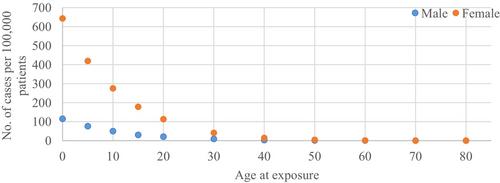
Although the mean thyroid organ dose (2.66 ± 1.03 mGy) and ED (1.6 ± 0.4 mSv) were slightly higher in females, these differences were not statistically significant compared to males (mean thyroid dose, 2.52 ± 1.31 mGy; mean ED, 1.5 ± 0.4 mSv). Conversely, there was a significant difference between the mean thyroid LAR of females (0.91 ± 1.35) and males (0.20136 ± 0.29) (P = 0.001). However, the influencing parameters were virtually identical for both groups.
Conclusions
The study's results indicate that females have a higher LAR than males, which can be attributed to higher radiation sensitivity of the thyroid in females. Thus, additional care in the choice of scan parameters and irradiated scan field for female patients is recommended.
期刊介绍:
Journal of Medical Radiation Sciences (JMRS) is an international and multidisciplinary peer-reviewed journal that accepts manuscripts related to medical imaging / diagnostic radiography, radiation therapy, nuclear medicine, medical ultrasound / sonography, and the complementary disciplines of medical physics, radiology, radiation oncology, nursing, psychology and sociology. Manuscripts may take the form of: original articles, review articles, commentary articles, technical evaluations, case series and case studies. JMRS promotes excellence in international medical radiation science by the publication of contemporary and advanced research that encourages the adoption of the best clinical, scientific and educational practices in international communities. JMRS is the official professional journal of the Australian Society of Medical Imaging and Radiation Therapy (ASMIRT) and the New Zealand Institute of Medical Radiation Technology (NZIMRT).