Risk indicators for gingival recession in the esthetic zone: A cross-sectional clinical, tomographic, and ultrasonographic study

IF 4.2 2区 医学 Q1 DENTISTRY, ORAL SURGERY & MEDICINE
Kathleen Chloe Mascardo, Justin Tomack, Chia-Yu Chen, Leonardo Mancini, David M. Kim, Bernard Friedland, Shayan Barootchi, Lorenzo Tavelli
{"title":"Risk indicators for gingival recession in the esthetic zone: A cross-sectional clinical, tomographic, and ultrasonographic study","authors":"Kathleen Chloe Mascardo,&nbsp;Justin Tomack,&nbsp;Chia-Yu Chen,&nbsp;Leonardo Mancini,&nbsp;David M. Kim,&nbsp;Bernard Friedland,&nbsp;Shayan Barootchi,&nbsp;Lorenzo Tavelli","doi":"10.1002/JPER.23-0357","DOIUrl":null,"url":null,"abstract":"<div>\n \n \n <section>\n \n <h3> Background</h3>\n \n <p>To evaluate the risk indicators associated with midfacial gingival recessions (GR) in the natural dentition esthetic regions.</p>\n </section>\n \n <section>\n \n <h3> Methods</h3>\n \n <p>Cone-beam computed tomography (CBCT) results of thirty-seven subjects presenting with 268 eligible teeth were included in the cross-sectional study. Clinical measurements included presence/absence of midfacial GR; the depth of the midfacial, mesial, and distal gingival recession; the recession type (RT); keratinized tissue width (KT); and attached gingiva width (AG). Questionnaires were utilized to capture patient-reported esthetics and dental hypersensitivity for each study tooth. Buccal bone dehiscence (cBBD) and buccal bone thickness (cBBT) were measured on the CBCT scans. High-frequency ultrasonography was performed to assess gingival thickness (GT) and buccal bone dehiscence (uBBD). Intraoral optical scanning was obtained to quantify the buccolingual position of each study site (3D profile analysis). Multilevel logistic regression analyses with generalized estimation equations were performed to assess the factors associated with the conditions of interest.</p>\n </section>\n \n <section>\n \n <h3> Results</h3>\n \n <p>The presence of midfacial GR was significantly associated with the history of periodontal treatment for pocket reduction (OR 7.99, <i>p</i> = 0.006), KT (OR 0.62, <i>p</i> &lt; 0.001), cBBD (OR 2.30, <i>p</i> = 0.015), GT 1.5 mm from the gingival margin (OR 0.18, <i>p</i> = 0.04) and 3D profile 1 mm from the gingival margin (OR 1.04, <i>p</i> = 0.001). The depth of midfacial GR was significantly correlated to previous history of periodontal treatment (OR 0.96, <i>p</i> = 0.001), KT (OR −0.18, <i>p</i> &lt; 0.001), presence of bone fenestration (OR 0.24, <i>p</i> = 0.044), and cBBD (OR 0.43, <i>p</i> &lt; 0.001). The depth of midfacial GR was also the only factor associated with patient-reported esthetics (OR −3.38, <i>p</i> = 0.022), while KT (OR 0.77, <i>p</i> = 0.018) and AG (OR 0.82, <i>p</i> = 0.047) were significantly correlated with patient-reported dental hypersensitivity.</p>\n </section>\n \n <section>\n \n <h3> Conclusions</h3>\n \n <p>Several risk indicators of midfacial and interproximal GR in the esthetic region were identified. The use of imaging technologies allowed for detection of parameters associated with the conditions of interest, and, therefore, their incorporation in future clinical studies is advocated. Ultrasonography could be preferred over CBCT for a noninvasive assessment of periodontal phenotype.</p>\n </section>\n </div>","PeriodicalId":16716,"journal":{"name":"Journal of periodontology","volume":"95 5","pages":"432-443"},"PeriodicalIF":4.2000,"publicationDate":"2024-01-09","publicationTypes":"Journal Article","fieldsOfStudy":null,"isOpenAccess":false,"openAccessPdf":"https://onlinelibrary.wiley.com/doi/epdf/10.1002/JPER.23-0357","citationCount":"0","resultStr":null,"platform":"Semanticscholar","paperid":null,"PeriodicalName":"Journal of periodontology","FirstCategoryId":"3","ListUrlMain":"https://onlinelibrary.wiley.com/doi/10.1002/JPER.23-0357","RegionNum":2,"RegionCategory":"医学","ArticlePicture":[],"TitleCN":null,"AbstractTextCN":null,"PMCID":null,"EPubDate":"","PubModel":"","JCR":"Q1","JCRName":"DENTISTRY, ORAL SURGERY & MEDICINE","Score":null,"Total":0}
引用次数: 0

Abstract

Background

To evaluate the risk indicators associated with midfacial gingival recessions (GR) in the natural dentition esthetic regions.

Methods

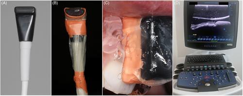
Cone-beam computed tomography (CBCT) results of thirty-seven subjects presenting with 268 eligible teeth were included in the cross-sectional study. Clinical measurements included presence/absence of midfacial GR; the depth of the midfacial, mesial, and distal gingival recession; the recession type (RT); keratinized tissue width (KT); and attached gingiva width (AG). Questionnaires were utilized to capture patient-reported esthetics and dental hypersensitivity for each study tooth. Buccal bone dehiscence (cBBD) and buccal bone thickness (cBBT) were measured on the CBCT scans. High-frequency ultrasonography was performed to assess gingival thickness (GT) and buccal bone dehiscence (uBBD). Intraoral optical scanning was obtained to quantify the buccolingual position of each study site (3D profile analysis). Multilevel logistic regression analyses with generalized estimation equations were performed to assess the factors associated with the conditions of interest.

Results

The presence of midfacial GR was significantly associated with the history of periodontal treatment for pocket reduction (OR 7.99, p = 0.006), KT (OR 0.62, p < 0.001), cBBD (OR 2.30, p = 0.015), GT 1.5 mm from the gingival margin (OR 0.18, p = 0.04) and 3D profile 1 mm from the gingival margin (OR 1.04, p = 0.001). The depth of midfacial GR was significantly correlated to previous history of periodontal treatment (OR 0.96, p = 0.001), KT (OR −0.18, p < 0.001), presence of bone fenestration (OR 0.24, p = 0.044), and cBBD (OR 0.43, p < 0.001). The depth of midfacial GR was also the only factor associated with patient-reported esthetics (OR −3.38, p = 0.022), while KT (OR 0.77, p = 0.018) and AG (OR 0.82, p = 0.047) were significantly correlated with patient-reported dental hypersensitivity.

Conclusions

Several risk indicators of midfacial and interproximal GR in the esthetic region were identified. The use of imaging technologies allowed for detection of parameters associated with the conditions of interest, and, therefore, their incorporation in future clinical studies is advocated. Ultrasonography could be preferred over CBCT for a noninvasive assessment of periodontal phenotype.
Abstract Image

美容区牙龈退缩的风险指标:一项横断面临床、断层和超声波研究。
背景:评估与天然牙美容区域中面部牙龈凹陷(GR)相关的风险指标:目的:评估与天然牙美容区面中部牙龈凹陷(GR)相关的风险指标:横断面研究纳入了 37 名受试者的锥形束计算机断层扫描(CBCT)结果,这些受试者有 268 颗符合条件的牙齿。临床测量包括是否存在面中部GR;面中部、中侧和远侧牙龈退缩的深度;退缩类型(RT);角化组织宽度(KT);附着龈宽度(AG)。利用调查问卷来了解患者对每颗研究牙齿的美观度和牙齿过敏性的报告。通过 CBCT 扫描测量颊骨开裂(cBBD)和颊骨厚度(cBBT)。高频超声波检查用于评估牙龈厚度(GT)和颊骨开裂(uBBD)。口内光学扫描用于量化每个研究部位的颊舌向位置(三维轮廓分析)。使用广义估计方程进行多层次逻辑回归分析,以评估与相关情况有关的因素:结果:中面部 GR 的存在与牙周治疗史(OR 7.99,p = 0.006)、KT(OR 0.62,p 结论:中面部 GR 与牙周治疗史有显著相关性:研究发现了美学区域面中部和近端间 GR 的几个风险指标。使用成像技术可以检测出与相关情况有关的参数,因此,建议将其纳入未来的临床研究中。在对牙周表型进行无创评估时,超声波成像技术可能比 CBCT 更受青睐。
本文章由计算机程序翻译,如有差异,请以英文原文为准。
求助全文
约1分钟内获得全文 求助全文
来源期刊
Journal of periodontology
Journal of periodontology 医学-牙科与口腔外科
CiteScore
9.10
自引率
7.00%
发文量
290
审稿时长
3-8 weeks
期刊介绍: The Journal of Periodontology publishes articles relevant to the science and practice of periodontics and related areas.
×
引用
GB/T 7714-2015
复制
MLA
复制
APA
复制
导出至
BibTeX EndNote RefMan NoteFirst NoteExpress
×
提示
您的信息不完整,为了账户安全,请先补充。
现在去补充
×
提示
您因"违规操作"
具体请查看互助需知
我知道了
×
提示
确定
请完成安全验证×
copy
已复制链接
快去分享给好友吧!
我知道了
右上角分享
点击右上角分享
0
联系我们:info@booksci.cn Book学术提供免费学术资源搜索服务,方便国内外学者检索中英文文献。致力于提供最便捷和优质的服务体验。 Copyright © 2023 布克学术 All rights reserved.
京ICP备2023020795号-1
ghs 京公网安备 11010802042870号
Book学术文献互助
Book学术文献互助群
群 号:481959085
Book学术官方微信