{"title":"<sup>18</sup>F-FDG PET/CT imaging of IgG4-producing MALT lymphoma with multiple site involvement.","authors":"Kodai Kawaji, Seiji Kurata, Katsuhisa Matsuo, Hiroaki Miyoshi, Jun Akiba, Fumihiko Mouri, Akiko Sumi, Kiminori Fujimoto, Toshi Abe","doi":"10.22038/AOJNMB.2023.73477.1512","DOIUrl":null,"url":null,"abstract":"<p><p><sup>18</sup>F-FDG PET/CT is regarded as a modality utilized for the purpose of lesion localization, staging and assessment of treatment response in patients with lymphoma. However, it is difficult that we diagnose among multifocal lymphoma, IgG4-related disease (IgG4-RD), or a combination of both conditions when confronted with multiple sites of <sup>18</sup>F-FDG uptake with heightened serum IgG4 levels. We present a case of a 72-year-old male who was suspected of Sjögren's syndrome based on symptoms of xerostomia accompanied by swelling of the bilateral upper eyelid and salivary glands. Following a diagnostic biopsy that revealed mucosa-associated lymphoid tissue (MALT) lymphoma as a possible finding, <sup>18</sup>F-FDG PET/CT was conducted, which demonstrated multiple sites of <sup>18</sup>F-FDG accumulation. While multifocal MALT lymphoma was initially suspected, the coexistence of IgG4-RD could not be definitively ruled out due to the elevated serum IgG4 levels. Subsequent histopathological and immunohistochemical examinations confirmed the diagnosis of IgG4-producing MALT lymphoma. After receiving systemic therapy with rituximab, the swelling of the bilateral upper eyelid and parotid glands resolved upon visual examination, and the serum IgG4 levels returned to within the normal range in a few months. No new lesions were detected during the subsequent follow-up examinations conducted over a period of 3 years.</p>","PeriodicalId":8503,"journal":{"name":"Asia Oceania Journal of Nuclear Medicine and Biology","volume":"12 1","pages":"52-56"},"PeriodicalIF":0.0000,"publicationDate":"2024-01-01","publicationTypes":"Journal Article","fieldsOfStudy":null,"isOpenAccess":false,"openAccessPdf":"https://www.ncbi.nlm.nih.gov/pmc/articles/PMC10757051/pdf/","citationCount":"0","resultStr":null,"platform":"Semanticscholar","paperid":null,"PeriodicalName":"Asia Oceania Journal of Nuclear Medicine and Biology","FirstCategoryId":"1085","ListUrlMain":"https://doi.org/10.22038/AOJNMB.2023.73477.1512","RegionNum":0,"RegionCategory":null,"ArticlePicture":[],"TitleCN":null,"AbstractTextCN":null,"PMCID":null,"EPubDate":"","PubModel":"","JCR":"Q3","JCRName":"Medicine","Score":null,"Total":0}
引用次数: 0
Abstract
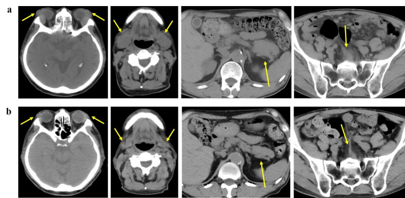
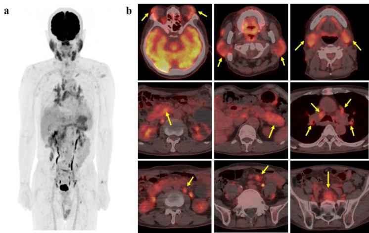
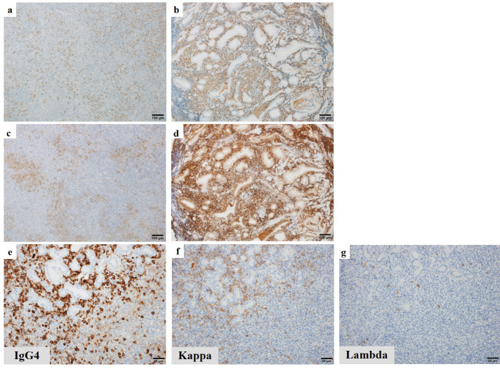
18F-FDG PET/CT is regarded as a modality utilized for the purpose of lesion localization, staging and assessment of treatment response in patients with lymphoma. However, it is difficult that we diagnose among multifocal lymphoma, IgG4-related disease (IgG4-RD), or a combination of both conditions when confronted with multiple sites of 18F-FDG uptake with heightened serum IgG4 levels. We present a case of a 72-year-old male who was suspected of Sjögren's syndrome based on symptoms of xerostomia accompanied by swelling of the bilateral upper eyelid and salivary glands. Following a diagnostic biopsy that revealed mucosa-associated lymphoid tissue (MALT) lymphoma as a possible finding, 18F-FDG PET/CT was conducted, which demonstrated multiple sites of 18F-FDG accumulation. While multifocal MALT lymphoma was initially suspected, the coexistence of IgG4-RD could not be definitively ruled out due to the elevated serum IgG4 levels. Subsequent histopathological and immunohistochemical examinations confirmed the diagnosis of IgG4-producing MALT lymphoma. After receiving systemic therapy with rituximab, the swelling of the bilateral upper eyelid and parotid glands resolved upon visual examination, and the serum IgG4 levels returned to within the normal range in a few months. No new lesions were detected during the subsequent follow-up examinations conducted over a period of 3 years.

 求助内容:
求助内容: 应助结果提醒方式:


