Arsenic Exposure Induces Neuro-immune Toxicity in the Cerebral Cortex and the Hippocampus via Neuroglia and NLRP3 Inflammasome Activation in C57BL/6 Mice.
IF 4.3 3区 材料科学Q1 ENGINEERING, ELECTRICAL & ELECTRONIC
Nan Yan, Zhengdong Wang, Zhou Li, Yang Zheng, Nan Chang, Kangjie Xu, Qian Wang, Xiaoxu Duan
{"title":"Arsenic Exposure Induces Neuro-immune Toxicity in the Cerebral Cortex and the Hippocampus via Neuroglia and NLRP3 Inflammasome Activation in C57BL/6 Mice.","authors":"Nan Yan, Zhengdong Wang, Zhou Li, Yang Zheng, Nan Chang, Kangjie Xu, Qian Wang, Xiaoxu Duan","doi":"10.1007/s12011-023-04012-4","DOIUrl":null,"url":null,"abstract":"<p><p>This study aimed to examine the immuntoxic effects of arsenic in the nervous system. Our results showed that arsenic increased corticocerebral and hippocampal weights (p < 0.05). Morris water maze tests revealed that arsenic significantly increased the time spent in latency to platform on the fourth day in 50 mg/L arsenic exposure and the fifth day in 25 and 50 mg/L arsenic exposure, as well as reduced the path length in target quadrant, time spent in target quadrant, and crossing times of the platform (p < 0.05). Hematoxylin-eosin staining showed that the vacuolated degeneration and pyknosis was found in the cerebral cortex and hippocampus of arsenic-treated mice. The mRNA levels of corticocerebral and hippocampal brain-derived neurotrophic factor (BDNF) and vascular endothelial growth factor (VEGF) were decreased in the 50 mg/L arsenic-treated group (p < 0.05). In addition, immunofluorescence staining showed that 25 and 50 mg/L arsenic all increased the expression of CD11b and glial fibrillary acidic protein (GFAP) in the cerebral cortex and hippocampus (p < 0.05). Arsenic markedly raised antigen-presenting molecule MHCII and CD40 mRNA levels in the cerebral cortex and hippocampus and upregulated the cell chemokine receptor 5 (CCR5) and CCR7 mRNA levels in the cerebral cortex at the 50 mg/L arsenic group, and increased the CCR7 mRNA levels in the hippocampus at the 25 and 50 mg/L arsenic groups (p < 0.05). Arsenic activated the nucleotide-binding domain-like receptor protein-3 (NLRP3) inflammasome, and enhanced its upstream promoter NF-κB protein level and downstream regulators IL-18 mRNA levels. Collectively, these results provide new evidences for the neuro-immune toxicity of arsenic.</p>","PeriodicalId":3,"journal":{"name":"ACS Applied Electronic Materials","volume":null,"pages":null},"PeriodicalIF":4.3000,"publicationDate":"2024-10-01","publicationTypes":"Journal Article","fieldsOfStudy":null,"isOpenAccess":false,"openAccessPdf":"","citationCount":"0","resultStr":null,"platform":"Semanticscholar","paperid":null,"PeriodicalName":"ACS Applied Electronic Materials","FirstCategoryId":"99","ListUrlMain":"https://doi.org/10.1007/s12011-023-04012-4","RegionNum":3,"RegionCategory":"材料科学","ArticlePicture":[],"TitleCN":null,"AbstractTextCN":null,"PMCID":null,"EPubDate":"2023/12/27 0:00:00","PubModel":"Epub","JCR":"Q1","JCRName":"ENGINEERING, ELECTRICAL & ELECTRONIC","Score":null,"Total":0}
引用次数: 0
Abstract
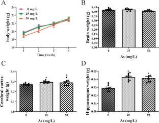
This study aimed to examine the immuntoxic effects of arsenic in the nervous system. Our results showed that arsenic increased corticocerebral and hippocampal weights (p < 0.05). Morris water maze tests revealed that arsenic significantly increased the time spent in latency to platform on the fourth day in 50 mg/L arsenic exposure and the fifth day in 25 and 50 mg/L arsenic exposure, as well as reduced the path length in target quadrant, time spent in target quadrant, and crossing times of the platform (p < 0.05). Hematoxylin-eosin staining showed that the vacuolated degeneration and pyknosis was found in the cerebral cortex and hippocampus of arsenic-treated mice. The mRNA levels of corticocerebral and hippocampal brain-derived neurotrophic factor (BDNF) and vascular endothelial growth factor (VEGF) were decreased in the 50 mg/L arsenic-treated group (p < 0.05). In addition, immunofluorescence staining showed that 25 and 50 mg/L arsenic all increased the expression of CD11b and glial fibrillary acidic protein (GFAP) in the cerebral cortex and hippocampus (p < 0.05). Arsenic markedly raised antigen-presenting molecule MHCII and CD40 mRNA levels in the cerebral cortex and hippocampus and upregulated the cell chemokine receptor 5 (CCR5) and CCR7 mRNA levels in the cerebral cortex at the 50 mg/L arsenic group, and increased the CCR7 mRNA levels in the hippocampus at the 25 and 50 mg/L arsenic groups (p < 0.05). Arsenic activated the nucleotide-binding domain-like receptor protein-3 (NLRP3) inflammasome, and enhanced its upstream promoter NF-κB protein level and downstream regulators IL-18 mRNA levels. Collectively, these results provide new evidences for the neuro-immune toxicity of arsenic.

 求助内容:
求助内容: 应助结果提醒方式:


