The chicken chorioallantoic membrane as a low-cost, high-throughput model for cancer imaging

Lydia M. Smith, Hannah E. Greenwood, Will E. Tyrrell, Richard S. Edwards, Vittorio de Santis, Friedrich Baark, George Firth, Muhammet Tanc, Samantha Y. A. Terry, Anne Herrmann, Richard Southworth, Timothy H. Witney
{"title":"The chicken chorioallantoic membrane as a low-cost, high-throughput model for cancer imaging","authors":"Lydia M. Smith, Hannah E. Greenwood, Will E. Tyrrell, Richard S. Edwards, Vittorio de Santis, Friedrich Baark, George Firth, Muhammet Tanc, Samantha Y. A. Terry, Anne Herrmann, Richard Southworth, Timothy H. Witney","doi":"10.1038/s44303-023-00001-3","DOIUrl":null,"url":null,"abstract":"Mouse models are invaluable tools for radiotracer development and validation. They are, however, expensive, low throughput, and are constrained by animal welfare considerations. Here, we assessed the chicken chorioallantoic membrane (CAM) as an alternative to mice for preclinical cancer imaging studies. NCI-H460 FLuc cells grown in Matrigel on the CAM formed vascularized tumors of reproducible size without compromising embryo viability. By designing a simple method for vessel cannulation it was possible to perform dynamic PET imaging in ovo, producing high tumor-to-background signal for both 18F-2-fluoro-2-deoxy-D-glucose (18F-FDG) and (4S)-4-(3-18F-fluoropropyl)-L-glutamate (18F-FSPG). The pattern of 18F-FDG tumor uptake were similar in ovo and in vivo, although tumor-associated radioactivity was higher in the CAM-grown tumors over the 60 min imaging time course. Additionally, 18F-FSPG provided an early marker of both treatment response to external beam radiotherapy and target inhibition in ovo. Overall, the CAM provided a low-cost alternative to tumor xenograft mouse models which may broaden access to PET and SPECT imaging and have utility across multiple applications.","PeriodicalId":501709,"journal":{"name":"npj Imaging","volume":" ","pages":"1-12"},"PeriodicalIF":0.0000,"publicationDate":"2023-11-29","publicationTypes":"Journal Article","fieldsOfStudy":null,"isOpenAccess":false,"openAccessPdf":"https://www.nature.com/articles/s44303-023-00001-3.pdf","citationCount":"0","resultStr":null,"platform":"Semanticscholar","paperid":null,"PeriodicalName":"npj Imaging","FirstCategoryId":"1085","ListUrlMain":"https://www.nature.com/articles/s44303-023-00001-3","RegionNum":0,"RegionCategory":null,"ArticlePicture":[],"TitleCN":null,"AbstractTextCN":null,"PMCID":null,"EPubDate":"","PubModel":"","JCR":"","JCRName":"","Score":null,"Total":0}
引用次数: 0

Abstract

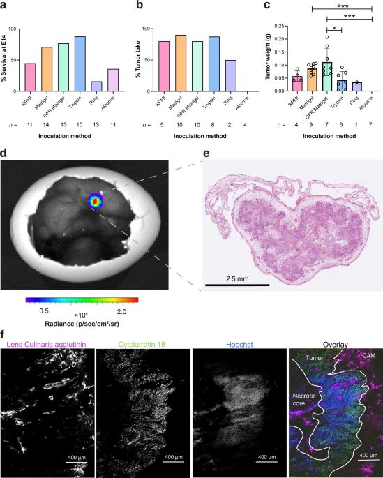
Mouse models are invaluable tools for radiotracer development and validation. They are, however, expensive, low throughput, and are constrained by animal welfare considerations. Here, we assessed the chicken chorioallantoic membrane (CAM) as an alternative to mice for preclinical cancer imaging studies. NCI-H460 FLuc cells grown in Matrigel on the CAM formed vascularized tumors of reproducible size without compromising embryo viability. By designing a simple method for vessel cannulation it was possible to perform dynamic PET imaging in ovo, producing high tumor-to-background signal for both 18F-2-fluoro-2-deoxy-D-glucose (18F-FDG) and (4S)-4-(3-18F-fluoropropyl)-L-glutamate (18F-FSPG). The pattern of 18F-FDG tumor uptake were similar in ovo and in vivo, although tumor-associated radioactivity was higher in the CAM-grown tumors over the 60 min imaging time course. Additionally, 18F-FSPG provided an early marker of both treatment response to external beam radiotherapy and target inhibition in ovo. Overall, the CAM provided a low-cost alternative to tumor xenograft mouse models which may broaden access to PET and SPECT imaging and have utility across multiple applications.
Abstract Image

鸡绒毛膜是一种低成本、高通量的癌症成像模型
小鼠模型是放射性示踪剂开发和验证的宝贵工具。然而,小鼠模型价格昂贵、通量低,而且受到动物福利因素的限制。在这里,我们评估了鸡绒毛膜(CAM)作为小鼠临床前癌症成像研究的替代品。在CAM上的Matrigel中生长的NCI-H460 FLuc细胞形成了大小可重复的血管化肿瘤,而不会影响胚胎的存活率。通过设计一种简单的血管插管方法,可以在胚胎中进行动态 PET 成像,为 18F-2-fluoro-2-deoxy-D-glucose (18F-FDG) 和 (4S)-4-(3-18F-fluoropropyl)-L-glutamate (18F-FSPG) 产生较高的肿瘤-背景信号。18F-FDG的肿瘤摄取模式在体内和体外相似,但在60分钟的成像过程中,CAM生长的肿瘤中肿瘤相关放射性更高。此外,18F-FSPG 还是体外放疗反应和体内靶点抑制的早期标记物。总之,CAM 为肿瘤异种移植小鼠模型提供了一种低成本的替代方法,它可以拓宽 PET 和 SPECT 成像的应用范围,并在多种应用中发挥作用。
本文章由计算机程序翻译,如有差异,请以英文原文为准。
求助全文
约1分钟内获得全文 求助全文
来源期刊
自引率
0.00%
发文量
0
×
引用
GB/T 7714-2015
复制
MLA
复制
APA
复制
导出至
BibTeX EndNote RefMan NoteFirst NoteExpress
×
提示
您的信息不完整,为了账户安全,请先补充。
现在去补充
×
提示
您因"违规操作"
具体请查看互助需知
我知道了
×
提示
确定
请完成安全验证×
copy
已复制链接
快去分享给好友吧!
我知道了
右上角分享
点击右上角分享
0
联系我们:info@booksci.cn Book学术提供免费学术资源搜索服务,方便国内外学者检索中英文文献。致力于提供最便捷和优质的服务体验。 Copyright © 2023 布克学术 All rights reserved.
京ICP备2023020795号-1
ghs 京公网安备 11010802042870号
Book学术文献互助
Book学术文献互助群
群 号:481959085
Book学术官方微信