Franziska Eckardt, Robin Mittas, Nastassya Horlava, Johannes Schiefelbein, Ben Asani, Stylianos Michalakis, Maximilian Gerhardt, Claudia Priglinger, Daniel Keeser, Nikolaos Koutsouleris, Siegfried Priglinger, Fabian Theis, Tingying Peng, Benedikt Schworm
{"title":"Deep Learning-Based Retinal Layer Segmentation in Optical Coherence Tomography Scans of Patients with Inherited Retinal Diseases.","authors":"Franziska Eckardt, Robin Mittas, Nastassya Horlava, Johannes Schiefelbein, Ben Asani, Stylianos Michalakis, Maximilian Gerhardt, Claudia Priglinger, Daniel Keeser, Nikolaos Koutsouleris, Siegfried Priglinger, Fabian Theis, Tingying Peng, Benedikt Schworm","doi":"10.1055/a-2227-3742","DOIUrl":null,"url":null,"abstract":"<p><strong>Background: </strong>In optical coherence tomography (OCT) scans of patients with inherited retinal diseases (IRDs), the measurement of the thickness of the outer nuclear layer (ONL) has been well established as a surrogate marker for photoreceptor preservation. Current automatic segmentation tools fail in OCT segmentation in IRDs, and manual segmentation is time-consuming.</p><p><strong>Methods and material: </strong>Patients with IRD and an available OCT scan were screened for the present study. Additionally, OCT scans of patients without retinal disease were included to provide training data for artificial intelligence (AI). We trained a U-net-based model on healthy patients and applied a domain adaption technique to the IRD patients' scans.</p><p><strong>Results: </strong>We established an AI-based image segmentation algorithm that reliably segments the ONL in OCT scans of IRD patients. In a test dataset, the dice score of the algorithm was 98.7%. Furthermore, we generated thickness maps of the full retinal thickness and the ONL layer for each patient.</p><p><strong>Conclusion: </strong>Accurate segmentation of anatomical layers on OCT scans plays a crucial role for predictive models linking retinal structure to visual function. Our algorithm for segmentation of OCT images could provide the basis for further studies on IRDs.</p>","PeriodicalId":17904,"journal":{"name":"Klinische Monatsblatter fur Augenheilkunde","volume":" ","pages":"903-911"},"PeriodicalIF":0.7000,"publicationDate":"2025-09-01","publicationTypes":"Journal Article","fieldsOfStudy":null,"isOpenAccess":false,"openAccessPdf":"https://www.ncbi.nlm.nih.gov/pmc/articles/PMC12445938/pdf/","citationCount":"0","resultStr":null,"platform":"Semanticscholar","paperid":null,"PeriodicalName":"Klinische Monatsblatter fur Augenheilkunde","FirstCategoryId":"3","ListUrlMain":"https://doi.org/10.1055/a-2227-3742","RegionNum":4,"RegionCategory":"医学","ArticlePicture":[],"TitleCN":null,"AbstractTextCN":null,"PMCID":null,"EPubDate":"2023/12/12 0:00:00","PubModel":"Epub","JCR":"Q4","JCRName":"OPHTHALMOLOGY","Score":null,"Total":0}
引用次数: 0
Abstract
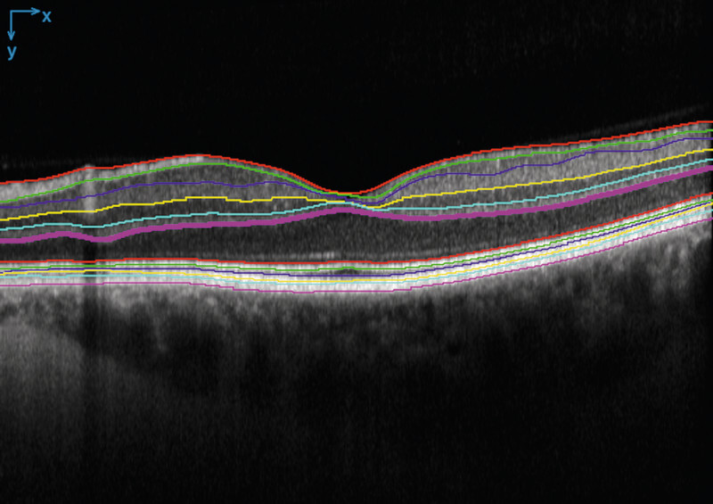
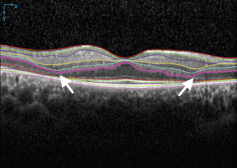
Background: In optical coherence tomography (OCT) scans of patients with inherited retinal diseases (IRDs), the measurement of the thickness of the outer nuclear layer (ONL) has been well established as a surrogate marker for photoreceptor preservation. Current automatic segmentation tools fail in OCT segmentation in IRDs, and manual segmentation is time-consuming.
Methods and material: Patients with IRD and an available OCT scan were screened for the present study. Additionally, OCT scans of patients without retinal disease were included to provide training data for artificial intelligence (AI). We trained a U-net-based model on healthy patients and applied a domain adaption technique to the IRD patients' scans.
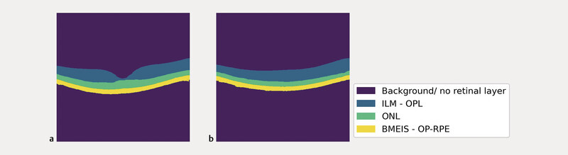
Results: We established an AI-based image segmentation algorithm that reliably segments the ONL in OCT scans of IRD patients. In a test dataset, the dice score of the algorithm was 98.7%. Furthermore, we generated thickness maps of the full retinal thickness and the ONL layer for each patient.
Conclusion: Accurate segmentation of anatomical layers on OCT scans plays a crucial role for predictive models linking retinal structure to visual function. Our algorithm for segmentation of OCT images could provide the basis for further studies on IRDs.
背景:在遗传性视网膜疾病(IRDs)患者的光学相干断层扫描(OCT)扫描中,核外层(ONL)厚度的测量已被公认为是感光细胞保存的替代标记。目前的自动分割工具无法对IRD进行OCT分割,而手动分割又非常耗时:本研究筛选了具有可用 OCT 扫描的 IRD 患者。此外,还包括无视网膜疾病患者的 OCT 扫描,以便为人工智能(AI)提供训练数据。我们在健康患者身上训练了一个基于 U-net 的模型,并对 IRD 患者的扫描应用了领域适应技术:结果:我们建立了一种基于人工智能的图像分割算法,它能可靠地分割 IRD 患者 OCT 扫描中的 ONL。在测试数据集中,该算法的骰子得分率为98.7%。此外,我们还为每位患者生成了全视网膜厚度图和 ONL 层厚度图:结论:准确分割 OCT 扫描图像上的解剖层对于建立视网膜结构与视觉功能相关联的预测模型至关重要。我们的 OCT 图像分割算法可为进一步研究 IRD 提供基础。
期刊介绍:
-Konzentriertes Fachwissen aus Klinik und Praxis:
Die entscheidenden Ergebnisse der internationalen Forschung - für Sie auf den Punkt gebracht und kritisch kommentiert,
Übersichtsarbeiten zu den maßgeblichen Themen der täglichen Praxis,
Top informiert - breite klinische Berichterstattung.
-CME-Punkte sammeln mit dem Refresher:
Effiziente, CME-zertifizierte Fortbildung, mit dem Refresher,
3 CME-Punkte pro Ausgabe - bis zu 36 CME-Punkte im Jahr!.
-Aktuelle Rubriken mit echtem Nutzwert:
Kurzreferate zu den wichtigsten Artikeln internationaler Zeitschriften,
Schwerpunktthema in jedem Heft: Ausführliche Übersichtsarbeiten zu den wichtigsten Themen der Ophthalmologie – so behalten Sie das gesamte Fach im Blick!,
Originalien mit den neuesten Entwicklungen,
Übersichten zu den relevanten Themen.

 求助内容:
求助内容: 应助结果提醒方式:


