K. V. Sergeeva, S. A. Tyganov, V. E. Kalashnikov, B. S. Shenkman, T. M. Mirzoev
{"title":"Role of Piezo1 Channels in Mechano-Anabolic Coupling in Rat Soleus Muscle","authors":"K. V. Sergeeva, S. A. Tyganov, V. E. Kalashnikov, B. S. Shenkman, T. M. Mirzoev","doi":"10.1134/S1990747823050082","DOIUrl":null,"url":null,"abstract":"<p>It is known that activation of protein synthesis and hypertrophy of muscle fibers in response to mechanical stress is realized through an anabolic mTORC1-dependent signaling pathway. However, mechanosensors through which a mechanical signal can be perceived and further transmitted to the mTORC1-dependent signaling pathway (mechanotransduction) are poorly identified. Mechanically activated (MA) ion channels are candidates for the role of such sarcolemmal mechanosensors. In this regard, the aim of this study was to investigate the potential role of MA channels (Piezo1) in the activation of the mTORC1-dependent pathway in the isolated rat soleus muscle in response to mechanical stress. Wistar rats were divided into 3 groups: (1) “Control” (animal muscles were not exposed to MA channel inhibitor or Piezo1 channel activator); (2) “Gadolinium” (animal muscles were incubated with gadolinium chloride, MA channel inhibitor), and (3) “Yoda” (animal muscles were incubated with Yoda1, Piezo1 MA channel activator). In rats from each group, <i>m. soleus</i> was isolated from the left limb and incubated in the appropriate solution without mechanical stress in the form of a series of stretching (“Rest”); <i>m. soleus</i> from the right limb was subjected to a series of stretching (“Stretch”) and then incubated in the appropriate solution. Phosphorylation of mTORC1 targets (p70S6K, rpS6, and 4E-BP1) in rat <i>m. soleus</i> was determined by PAAG electrophoresis and immunoblotting. A series of passive stretches of isolated <i>m. soleus</i> led to an increase in the phosphorylation of p70S6K, its substrate rpS6, and 4E-BP1 by 38.5, 168 and 112%, respectively, compared to the muscle that was not subjected to mechanical stress. Incubation of the muscles with gadolinium completely prevented the activation of mTORC1 markers caused by a series of stretches. Incubation of <i>m. soleus</i> in a solution with Yoda1 resulted in a decrease in the mechano-dependent phosphorylation of p70S6K, rpS6 and 4E-BP1 compared to the muscle that was not exposed to Yoda1. Thus, the methodological approach used in this work did not reveal the participation of Piezo1 in mechano-anabolic coupling in rat <i>m. soleus</i>.</p>","PeriodicalId":484,"journal":{"name":"Biochemistry (Moscow), Supplement Series A: Membrane and Cell Biology","volume":"17 4","pages":"286 - 292"},"PeriodicalIF":1.1000,"publicationDate":"2023-12-10","publicationTypes":"Journal Article","fieldsOfStudy":null,"isOpenAccess":false,"openAccessPdf":"","citationCount":"0","resultStr":null,"platform":"Semanticscholar","paperid":null,"PeriodicalName":"Biochemistry (Moscow), Supplement Series A: Membrane and Cell Biology","FirstCategoryId":"2","ListUrlMain":"https://link.springer.com/article/10.1134/S1990747823050082","RegionNum":0,"RegionCategory":null,"ArticlePicture":[],"TitleCN":null,"AbstractTextCN":null,"PMCID":null,"EPubDate":"","PubModel":"","JCR":"Q4","JCRName":"CELL BIOLOGY","Score":null,"Total":0}
引用次数: 0
Abstract
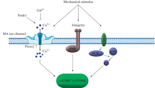
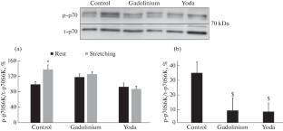
It is known that activation of protein synthesis and hypertrophy of muscle fibers in response to mechanical stress is realized through an anabolic mTORC1-dependent signaling pathway. However, mechanosensors through which a mechanical signal can be perceived and further transmitted to the mTORC1-dependent signaling pathway (mechanotransduction) are poorly identified. Mechanically activated (MA) ion channels are candidates for the role of such sarcolemmal mechanosensors. In this regard, the aim of this study was to investigate the potential role of MA channels (Piezo1) in the activation of the mTORC1-dependent pathway in the isolated rat soleus muscle in response to mechanical stress. Wistar rats were divided into 3 groups: (1) “Control” (animal muscles were not exposed to MA channel inhibitor or Piezo1 channel activator); (2) “Gadolinium” (animal muscles were incubated with gadolinium chloride, MA channel inhibitor), and (3) “Yoda” (animal muscles were incubated with Yoda1, Piezo1 MA channel activator). In rats from each group, m. soleus was isolated from the left limb and incubated in the appropriate solution without mechanical stress in the form of a series of stretching (“Rest”); m. soleus from the right limb was subjected to a series of stretching (“Stretch”) and then incubated in the appropriate solution. Phosphorylation of mTORC1 targets (p70S6K, rpS6, and 4E-BP1) in rat m. soleus was determined by PAAG electrophoresis and immunoblotting. A series of passive stretches of isolated m. soleus led to an increase in the phosphorylation of p70S6K, its substrate rpS6, and 4E-BP1 by 38.5, 168 and 112%, respectively, compared to the muscle that was not subjected to mechanical stress. Incubation of the muscles with gadolinium completely prevented the activation of mTORC1 markers caused by a series of stretches. Incubation of m. soleus in a solution with Yoda1 resulted in a decrease in the mechano-dependent phosphorylation of p70S6K, rpS6 and 4E-BP1 compared to the muscle that was not exposed to Yoda1. Thus, the methodological approach used in this work did not reveal the participation of Piezo1 in mechano-anabolic coupling in rat m. soleus.
期刊介绍:
Biochemistry (Moscow), Supplement Series A: Membrane and Cell Biology is an international peer reviewed journal that publishes original articles on physical, chemical, and molecular mechanisms that underlie basic properties of biological membranes and mediate membrane-related cellular functions. The primary topics of the journal are membrane structure, mechanisms of membrane transport, bioenergetics and photobiology, intracellular signaling as well as membrane aspects of cell biology, immunology, and medicine. The journal is multidisciplinary and gives preference to those articles that employ a variety of experimental approaches, basically in biophysics but also in biochemistry, cytology, and molecular biology. The journal publishes articles that strive for unveiling membrane and cellular functions through innovative theoretical models and computer simulations.
 求助内容:
求助内容: 应助结果提醒方式:


