A. A. Gaidash, V. K. Krut’ko, A. I. Kulak, O. N. Musskaya, K. V. Skrotskaya, Yu. P. Tokalchik, V. A. Kulchitsky
{"title":"Structure of the Peritenons of the Paravertebral Tendons Treated with Hyaluronic Acid","authors":"A. A. Gaidash, V. K. Krut’ko, A. I. Kulak, O. N. Musskaya, K. V. Skrotskaya, Yu. P. Tokalchik, V. A. Kulchitsky","doi":"10.1134/s2079086423060075","DOIUrl":null,"url":null,"abstract":"<h3 data-test=\"abstract-sub-heading\">Abstract</h3><p>The tendon sheaths (peritenons) of the paravertebral tendons of the tails of Wistar rats were studied using scanning electron microscopy. A phenomenological classification of the osteoid structures of the peritenons is given, with the identification of their persistent and permanent varieties. Sesamoid islets, needle-like and lamellar growths, and rudiments of osteons are classified as persistent. Persistent osteoid structures are well prepared for transformations aimed at strengthening the intracellular matrix under mechanical stress. Permanent osteoid structures are microgranules and faceted deposits of calcium phosphates involved in structural and mechanical processes and hetero- and homogeneous nucleation. Hyaluronate loosens the matrix of sesamoid islets, which increases the mobility of sesamoid globules and creates the prerequisites for their directed migration to areas of increased mechanical stress and foci of possible mineralization of extracellular substance, including fibrillar collagen. Hyaluronate sticks together granules and deposits of structured calcium phosphates and contributes to their growth and fixation in areas of increased risk of mechanical stress. This is a fundamentally important adaptive mechanism for strengthening the tendon tissue, acting in advance.</p>","PeriodicalId":9047,"journal":{"name":"Biology Bulletin Reviews","volume":"12 1","pages":""},"PeriodicalIF":0.0000,"publicationDate":"2023-11-28","publicationTypes":"Journal Article","fieldsOfStudy":null,"isOpenAccess":false,"openAccessPdf":"","citationCount":"0","resultStr":null,"platform":"Semanticscholar","paperid":null,"PeriodicalName":"Biology Bulletin Reviews","FirstCategoryId":"1085","ListUrlMain":"https://doi.org/10.1134/s2079086423060075","RegionNum":0,"RegionCategory":null,"ArticlePicture":[],"TitleCN":null,"AbstractTextCN":null,"PMCID":null,"EPubDate":"","PubModel":"","JCR":"","JCRName":"","Score":null,"Total":0}
引用次数: 0
Abstract
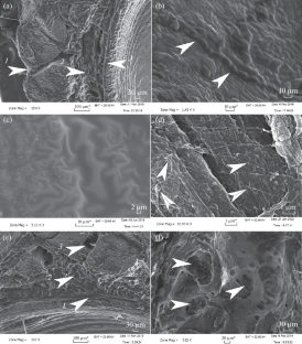
The tendon sheaths (peritenons) of the paravertebral tendons of the tails of Wistar rats were studied using scanning electron microscopy. A phenomenological classification of the osteoid structures of the peritenons is given, with the identification of their persistent and permanent varieties. Sesamoid islets, needle-like and lamellar growths, and rudiments of osteons are classified as persistent. Persistent osteoid structures are well prepared for transformations aimed at strengthening the intracellular matrix under mechanical stress. Permanent osteoid structures are microgranules and faceted deposits of calcium phosphates involved in structural and mechanical processes and hetero- and homogeneous nucleation. Hyaluronate loosens the matrix of sesamoid islets, which increases the mobility of sesamoid globules and creates the prerequisites for their directed migration to areas of increased mechanical stress and foci of possible mineralization of extracellular substance, including fibrillar collagen. Hyaluronate sticks together granules and deposits of structured calcium phosphates and contributes to their growth and fixation in areas of increased risk of mechanical stress. This is a fundamentally important adaptive mechanism for strengthening the tendon tissue, acting in advance.

 求助内容:
求助内容: 应助结果提醒方式:


