BCG infection dose guides dendritic cell migration and T cell priming in the draining lymph node

IF 4.1 4区 医学 Q2 IMMUNOLOGY
Veronika Krmeská, Lei Shen, Susanne Nylén, Pryscilla Fanini Wowk, Antonio Gigliotti Rothfuchs
{"title":"BCG infection dose guides dendritic cell migration and T cell priming in the draining lymph node","authors":"Veronika Krmeská, Lei Shen, Susanne Nylén, Pryscilla Fanini Wowk, Antonio Gigliotti Rothfuchs","doi":"10.1111/sji.13342","DOIUrl":null,"url":null,"abstract":"In contrast to delayed-type hypersensitivity (DTH) and other hallmark reactions of cell-mediated immunity that correlate with vaccine-mediated protection against <i>Mycobacterium tuberculosis</i>, the contribution of vaccine dose on responses that emerge early after infection in the skin with Bacille Calmette–Guérin (BCG) is not well understood. We used a mouse model of BCG skin infection to study the effect of BCG dose on the relocation of skin Dendritic cells (DCs) to draining lymph node (DLN). <i>Mycobacterium</i> antigen 85B-specific CD4<sup>+</sup> P25 T cell-receptor transgenic (P25 TCRTg) cells were used to probe priming to BCG in DLN. DC migration and T cell priming were studied across BCG inocula that varied up to 100-fold (10<sup>4</sup> to 10<sup>6</sup> Colony-forming units—CFUs). In line with earlier results in guinea pigs, DTH reaction in our model correlated with BCG dose. Importantly, priming of P25 TCRTg cells in DLN also escalated in a dose-dependent manner, peaking at day 6 after infection. Similar dose-escalation effects were seen for DC migration from infected skin and the accompanying transport of BCG to the DLN. BCG-triggered upregulation of co-stimulatory molecules on migratory DCs was restricted to the first 24 hour after infection and was independent of BCG dose over a 10-fold range (10<sup>5</sup> to 10<sup>6</sup> CFUs). The dose seemed to be a determinant of the number of total skin DCs that move to the DLN. In summary, our results support the use of higher BCG doses to detect robust DC migration and T cell priming.","PeriodicalId":21493,"journal":{"name":"Scandinavian Journal of Immunology","volume":null,"pages":null},"PeriodicalIF":4.1000,"publicationDate":"2023-11-21","publicationTypes":"Journal Article","fieldsOfStudy":null,"isOpenAccess":false,"openAccessPdf":"","citationCount":"0","resultStr":null,"platform":"Semanticscholar","paperid":null,"PeriodicalName":"Scandinavian Journal of Immunology","FirstCategoryId":"3","ListUrlMain":"https://doi.org/10.1111/sji.13342","RegionNum":4,"RegionCategory":"医学","ArticlePicture":[],"TitleCN":null,"AbstractTextCN":null,"PMCID":null,"EPubDate":"","PubModel":"","JCR":"Q2","JCRName":"IMMUNOLOGY","Score":null,"Total":0}
引用次数: 0

Abstract

In contrast to delayed-type hypersensitivity (DTH) and other hallmark reactions of cell-mediated immunity that correlate with vaccine-mediated protection against Mycobacterium tuberculosis, the contribution of vaccine dose on responses that emerge early after infection in the skin with Bacille Calmette–Guérin (BCG) is not well understood. We used a mouse model of BCG skin infection to study the effect of BCG dose on the relocation of skin Dendritic cells (DCs) to draining lymph node (DLN). Mycobacterium antigen 85B-specific CD4+ P25 T cell-receptor transgenic (P25 TCRTg) cells were used to probe priming to BCG in DLN. DC migration and T cell priming were studied across BCG inocula that varied up to 100-fold (104 to 106 Colony-forming units—CFUs). In line with earlier results in guinea pigs, DTH reaction in our model correlated with BCG dose. Importantly, priming of P25 TCRTg cells in DLN also escalated in a dose-dependent manner, peaking at day 6 after infection. Similar dose-escalation effects were seen for DC migration from infected skin and the accompanying transport of BCG to the DLN. BCG-triggered upregulation of co-stimulatory molecules on migratory DCs was restricted to the first 24 hour after infection and was independent of BCG dose over a 10-fold range (105 to 106 CFUs). The dose seemed to be a determinant of the number of total skin DCs that move to the DLN. In summary, our results support the use of higher BCG doses to detect robust DC migration and T cell priming.
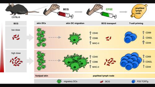
Abstract Image

卡介苗感染剂量引导引流淋巴结的树突状细胞迁移和T细胞启动
与延迟型超敏反应(DTH)和与疫苗介导的抗结核分枝杆菌保护相关的细胞介导免疫的其他标志性反应相反,疫苗剂量对皮肤感染卡介苗(BCG)后早期出现的反应的贡献尚不清楚。采用小鼠卡介苗皮肤感染模型,研究卡介苗剂量对皮肤树突状细胞(dc)向引流淋巴结(DLN)迁移的影响。采用分枝杆菌抗原85b特异性CD4+ P25 T细胞受体转基因细胞(P25 TCRTg)对DLN中BCG的引物进行检测。研究了DC迁移和T细胞启动在BCG接种中变化高达100倍(104至106集落形成单位- cfu)。与早期豚鼠的结果一致,我们模型中的DTH反应与卡介苗剂量相关。重要的是,P25 TCRTg细胞在DLN中的启动也以剂量依赖的方式增加,在感染后第6天达到峰值。DC从感染皮肤的迁移和伴随的卡介苗向DLN的运输也出现了类似的剂量递增效应。BCG触发的共刺激分子对迁移性dc的上调仅限于感染后的前24小时,并且在10倍范围内(105 ~ 106 cfu)与BCG剂量无关。剂量似乎是移动到DLN的总皮肤dc数量的决定因素。总之,我们的结果支持使用更高剂量的卡介苗来检测强大的DC迁移和T细胞启动。
本文章由计算机程序翻译,如有差异,请以英文原文为准。
求助全文
约1分钟内获得全文 求助全文
来源期刊
CiteScore
7.70
自引率
5.40%
发文量
109
审稿时长
1 months
期刊介绍: This peer-reviewed international journal publishes original articles and reviews on all aspects of basic, translational and clinical immunology. The journal aims to provide high quality service to authors, and high quality articles for readers. The journal accepts for publication material from investigators all over the world, which makes a significant contribution to basic, translational and clinical immunology.
×
引用
GB/T 7714-2015
复制
MLA
复制
APA
复制
导出至
BibTeX EndNote RefMan NoteFirst NoteExpress
×
提示
您的信息不完整,为了账户安全,请先补充。
现在去补充
×
提示
您因"违规操作"
具体请查看互助需知
我知道了
×
提示
确定
请完成安全验证×
copy
已复制链接
快去分享给好友吧!
我知道了
右上角分享
点击右上角分享
0
联系我们:info@booksci.cn Book学术提供免费学术资源搜索服务,方便国内外学者检索中英文文献。致力于提供最便捷和优质的服务体验。 Copyright © 2023 布克学术 All rights reserved.
京ICP备2023020795号-1
ghs 京公网安备 11010802042870号
Book学术文献互助
Book学术文献互助群
群 号:481959085
Book学术官方微信