{"title":"Automated Tractography for the Assessment of Aphasia in Acute Care Stroke Rehabilitation: A Case Series.","authors":"Midori Mochizuki, Yuki Uchiyama, Kazuhisa Domen, Tetsuo Koyama","doi":"10.2490/prm.20230041","DOIUrl":null,"url":null,"abstract":"<p><strong>Background: </strong>Aphasia is a common disorder among stroke patients. Assessment of aphasia is essential for scheduling appropriate rehabilitative treatment. Although this is conventionally accomplished using neuropsychological test batteries, these tests are not always accessible because of attention and/or consciousness disturbances during acute care. To overcome this issue, we have introduced a newly developed automated tractography known as XTRACT.</p><p><strong>Cases: </strong>Diffusion-tensor images were acquired from three patients on days 10-14. Brain images were processed by XTRACT, which automatically extracts neural tracts using standardized protocols. Fractional anisotropy (FA) values were then bilaterally evaluated in the following neural tracts associated with aphasia: arcuate fasciculus, inferior fronto-occipital fasciculus, middle longitudinal fasciculus, inferior longitudinal fasciculus, and uncinate fasciculus. Case 1 had word-finding difficulty on admission. FA values in the lesioned left hemisphere were not decreased in all tracts and this patient fully recovered during acute care. Case 2 had reduced spontaneous speech and a low FA value in the left arcuate fasciculus. Rehabilitative treatment was scheduled to improve the verbal output of sentences and word recall. Case 3 could not complete the conventional aphasia test battery because of attention disturbance. He had low FA values in all tracts in the left hemisphere. Rehabilitative treatment was designed to focus on both speaking and auditory comprehension.</p><p><strong>Discussion: </strong>Automated tractography enables quantitative assessment of the neural damage associated with aphasia, even in patients with attention and/or consciousness disturbances. This modality can aid in the assessment of aphasia and allows the planning of appropriate rehabilitative treatment.</p>","PeriodicalId":74584,"journal":{"name":"Progress in rehabilitation medicine","volume":"8 ","pages":"20230041"},"PeriodicalIF":1.5000,"publicationDate":"2023-11-22","publicationTypes":"Journal Article","fieldsOfStudy":null,"isOpenAccess":false,"openAccessPdf":"https://www.ncbi.nlm.nih.gov/pmc/articles/PMC10661235/pdf/","citationCount":"0","resultStr":null,"platform":"Semanticscholar","paperid":null,"PeriodicalName":"Progress in rehabilitation medicine","FirstCategoryId":"1085","ListUrlMain":"https://doi.org/10.2490/prm.20230041","RegionNum":0,"RegionCategory":null,"ArticlePicture":[],"TitleCN":null,"AbstractTextCN":null,"PMCID":null,"EPubDate":"2023/1/1 0:00:00","PubModel":"eCollection","JCR":"","JCRName":"","Score":null,"Total":0}
引用次数: 0
Abstract
Background: Aphasia is a common disorder among stroke patients. Assessment of aphasia is essential for scheduling appropriate rehabilitative treatment. Although this is conventionally accomplished using neuropsychological test batteries, these tests are not always accessible because of attention and/or consciousness disturbances during acute care. To overcome this issue, we have introduced a newly developed automated tractography known as XTRACT.
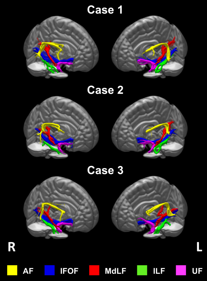
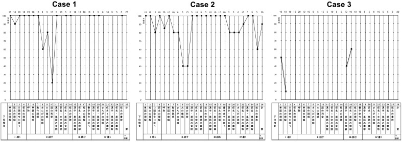
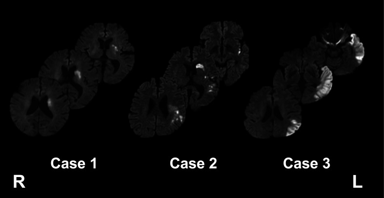
Cases: Diffusion-tensor images were acquired from three patients on days 10-14. Brain images were processed by XTRACT, which automatically extracts neural tracts using standardized protocols. Fractional anisotropy (FA) values were then bilaterally evaluated in the following neural tracts associated with aphasia: arcuate fasciculus, inferior fronto-occipital fasciculus, middle longitudinal fasciculus, inferior longitudinal fasciculus, and uncinate fasciculus. Case 1 had word-finding difficulty on admission. FA values in the lesioned left hemisphere were not decreased in all tracts and this patient fully recovered during acute care. Case 2 had reduced spontaneous speech and a low FA value in the left arcuate fasciculus. Rehabilitative treatment was scheduled to improve the verbal output of sentences and word recall. Case 3 could not complete the conventional aphasia test battery because of attention disturbance. He had low FA values in all tracts in the left hemisphere. Rehabilitative treatment was designed to focus on both speaking and auditory comprehension.
Discussion: Automated tractography enables quantitative assessment of the neural damage associated with aphasia, even in patients with attention and/or consciousness disturbances. This modality can aid in the assessment of aphasia and allows the planning of appropriate rehabilitative treatment.

 求助内容:
求助内容: 应助结果提醒方式:


