Gallbladder adenocarcinoma skin metastasis.
Q4 Medicine
Autopsy and Case Reports
Pub Date : 2023-11-27
eCollection Date: 2023-01-01
DOI:10.4322/acr.2023.458
引用次数: 0
Abstract
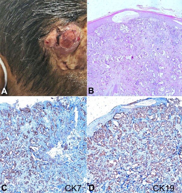
moderate cytoplasm. A few cells showed cytoplasmic clearing. The peritumoral desmoplastic reaction was also identified with intratumorally brisk mitotic figures. Focal areas of necrosis with acute and chronic inflammatory infiltrate were also identified. Immunohistochemistry revealed tumor cells to be diffusely positive for cytokeratin 7 (CK7) and CK19 (Figure 1C and 1D) and negative for CK20, thyroid transcription factor (TTF-1), Gross Cystic Disease Fluid Protein-15 (GCDFP-15), mammaglobin, Hepatocyte Paraffin 1 (Hep Par1), Paired box gene 8 (PAX-8) and Wilms tumor gene 1 (WT1). An abdominal computed tomography scan on imaging workup revealed a gallbladder mass with multiple liver lesions and brain metastasis. Based on immunohistochemistry and imaging findings, the diagnosis was metastatic gall bladder carcinoma to the scalp.
胆囊腺癌皮肤转移。
本文章由计算机程序翻译,如有差异,请以英文原文为准。
求助全文
约1分钟内获得全文
求助全文
来源期刊

Autopsy and Case Reports
Medicine-Internal Medicine
CiteScore
1.20
自引率
0.00%
发文量
60
审稿时长
9 weeks
 求助内容:
求助内容: 应助结果提醒方式:


