Immunophenotypic portrait of leukemia-associated-phenotype markers in B acute lymphoblastic leukemia
Abstract
Background
Multiparametric flow cytometry (MFC) is an essential diagnostic tool in B acute lymphoblastic leukemia (B ALL) to determine the B-lineage affiliation of the blast population and to define their complete immunophenotypic profile. Most MFC strategies used in routine laboratories include leukemia-associated phenotype (LAP) markers, whose expression profiles can be difficult to interpret. The aim of our study was to reach a better understanding of 7 LAP markers' landscape in B ALL: CD9, CD21, CD66c, CD58, CD81, CD123, and NG2.
Methods
Using a 10-color MFC approach, we evaluated the level of expression of 7 LAP markers including CD9, CD21, CD66c, CD58, CD81, CD123, and NG2, at the surface of normal peripheral blood leukocytes (n = 10 healthy donors), of normal precursor B regenerative cells (n = 40 uninvolved bone marrow samples) and of lymphoblasts (n = 100 peripheral blood samples or bone marrow samples from B ALL patients at diagnosis). The expression profile of B lymphoblasts was analyzed according the presence or absence of recurrent cytogenetic aberrations. The prognostic value of the 7 LAP markers was examined using Maxstat R algorithm.
Results
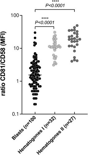
In order to help the interpretation of the MFC data in routine laboratories, we first determined internal positive and negative populations among normal leukocytes for each of the seven evaluated LAP markers. Second, their profile of expression was evaluated in normal B cell differentiation in comparison with B lymphoblasts to establish a synopsis of their expression in normal hematogones. We then evaluated the frequency of expression of these LAP markers at the surface of B lymphoblasts at diagnosis of B ALL. CD9 was expressed in 60% of the cases, CD21 in only 3% of the cases, CD58 in 96% of the cases, CD66c in 45% of the cases, CD81 in 97% of the cases, CD123 in 72% of the cases, and NG2 in only 2% of the cases. We confirmed the interest of the CD81/CD58 MFI expression ratio as a way to discriminate hematogones from lymphoblasts. We observed a significant lower expression of CD9 and of CD81 at the surface of B lymphoblasts with a t(9;22)(BCR-ABL) in comparison with B lymphoblasts without any recurrent cytogenetic alteration (p = 0.0317 and p = 0.0011, respectively) and with B lymphoblasts harboring other cytogenetic recurrent abnormalities (p = 0.0032 and p < 0.0001, respectively). B lymphoblasts with t(1;19) at diagnosis significantly overexpressed CD81 when compared with B lymphoblasts with other recurrent cytogenetic abnormalities or without any recurrent alteration (p = 0.0001). An overexpression of CD58 was also observed in the cases harboring this abnormal cytogenetic event, when compared with B lymphoblasts with other recurrent cytogenetic abnormalities (p = 0.030), or without any recurrent alteration (p = 0.0002). In addition, a high expression of CD123, of CD58 and of CD81 was associated with a favorable prognosis in our cohort of pediatric and young adult B ALL patients. We finally built a risk score based on the expression of these 3 LAP markers, this scoring approach being able to split these patients into a high-risk group (17%) and a better outcome group (83%, p < 0.0001).
Conclusion
The complexity of the phenotypic signature of lymphoblasts at diagnosis of B ALL is illustrated by the variability in the expression of LAP antigens. Knowledge of the expression levels of these markers in normal leukocytes and during normal B differentiation is crucial for an optimal interpretation of diagnostic cytometry results and serves as a basis for the biological follow-up of B ALL.
 求助内容:
求助内容: 应助结果提醒方式:


