Kevin Deschamps, Karel Mercken, Pieter Verschuren, Maarten Eerdekens, Eline Vanstraelen, Sander Wuite, Giovanni A Matricali
{"title":"Foot biomechanics in patients with advanced subtalar- and mid-tarsal joint osteoarthritis and poorly responding to conservative treatment.","authors":"Kevin Deschamps, Karel Mercken, Pieter Verschuren, Maarten Eerdekens, Eline Vanstraelen, Sander Wuite, Giovanni A Matricali","doi":"10.1186/s13047-023-00689-x","DOIUrl":null,"url":null,"abstract":"<p><strong>Background: </strong>A comprehensive insight into the effects of subtalar- and mid-tarsal joint osteoarthritis on lower limb's biomechanical characteristics during walking is lacking. Our goal was to assess joint kinematics and kinetics and compensatory mechanisms in patients with subtalar and mid-tarsal joint osteoarthritis.</p><p><strong>Methods: </strong>Patients with symptomatic and radiographically confirmed osteoarthritis of the subtalar and mid-tarsal (n = 10) and an asymptomatic control group (n = 10) were compared. Foot joint kinematics and kinetics during the stance phase of walking were quantified using a four-segment foot model.</p><p><strong>Results: </strong>During pre-swing phase, the tibio-talar range of motion in the sagittal plane of the patient group decreased significantly (P = 0.001), whereas the tarso-metatarsal joint range of motion in the sagittal plane was greater in the pre-swing phase (P = 0.003). The mid-tarsal joint showed lower transverse plane range of motion in the patient group during the loading response and pre-swing phase (P < 0.001 resp. P = 0.002). The patient group showed a lower Tibio-talar joint peak plantarflexion moment (P = 0.004), peak plantarflexion velocity (P < 0.001) and peak power generation in the sagittal plane (P < 0.001), and a lower mid-tarsal joint peak adduction and abduction velocity (P < 0.001 resp. P < 0.001) and peak power absorption (P < 0.001).</p><p><strong>Conclusions: </strong>These findings suggest that patients with subtalar and mid-tarsal joint osteoarthritis adopt a cautious walking strategy potentially dictated by pain, muscle weakness, kinesiophobia and stiffness. Since this poorly responding population faces surgical intervention on the short term, we recommend careful follow-up after fusion surgery since biomechanical outcome measures associated to this post-surgical stage is lacking.</p>","PeriodicalId":49164,"journal":{"name":"Journal of Foot and Ankle Research","volume":"16 1","pages":"85"},"PeriodicalIF":2.2000,"publicationDate":"2023-11-28","publicationTypes":"Journal Article","fieldsOfStudy":null,"isOpenAccess":false,"openAccessPdf":"https://www.ncbi.nlm.nih.gov/pmc/articles/PMC10683126/pdf/","citationCount":"0","resultStr":null,"platform":"Semanticscholar","paperid":null,"PeriodicalName":"Journal of Foot and Ankle Research","FirstCategoryId":"3","ListUrlMain":"https://doi.org/10.1186/s13047-023-00689-x","RegionNum":3,"RegionCategory":"医学","ArticlePicture":[],"TitleCN":null,"AbstractTextCN":null,"PMCID":null,"EPubDate":"","PubModel":"","JCR":"Q1","JCRName":"ORTHOPEDICS","Score":null,"Total":0}
引用次数: 0
Abstract
Background: A comprehensive insight into the effects of subtalar- and mid-tarsal joint osteoarthritis on lower limb's biomechanical characteristics during walking is lacking. Our goal was to assess joint kinematics and kinetics and compensatory mechanisms in patients with subtalar and mid-tarsal joint osteoarthritis.
Methods: Patients with symptomatic and radiographically confirmed osteoarthritis of the subtalar and mid-tarsal (n = 10) and an asymptomatic control group (n = 10) were compared. Foot joint kinematics and kinetics during the stance phase of walking were quantified using a four-segment foot model.
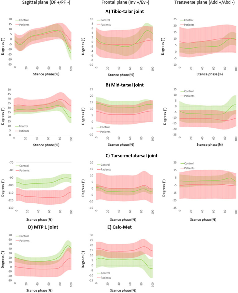
Results: During pre-swing phase, the tibio-talar range of motion in the sagittal plane of the patient group decreased significantly (P = 0.001), whereas the tarso-metatarsal joint range of motion in the sagittal plane was greater in the pre-swing phase (P = 0.003). The mid-tarsal joint showed lower transverse plane range of motion in the patient group during the loading response and pre-swing phase (P < 0.001 resp. P = 0.002). The patient group showed a lower Tibio-talar joint peak plantarflexion moment (P = 0.004), peak plantarflexion velocity (P < 0.001) and peak power generation in the sagittal plane (P < 0.001), and a lower mid-tarsal joint peak adduction and abduction velocity (P < 0.001 resp. P < 0.001) and peak power absorption (P < 0.001).
Conclusions: These findings suggest that patients with subtalar and mid-tarsal joint osteoarthritis adopt a cautious walking strategy potentially dictated by pain, muscle weakness, kinesiophobia and stiffness. Since this poorly responding population faces surgical intervention on the short term, we recommend careful follow-up after fusion surgery since biomechanical outcome measures associated to this post-surgical stage is lacking.
期刊介绍:
Journal of Foot and Ankle Research, the official journal of the Australian Podiatry Association and The College of Podiatry (UK), is an open access journal that encompasses all aspects of policy, organisation, delivery and clinical practice related to the assessment, diagnosis, prevention and management of foot and ankle disorders.
Journal of Foot and Ankle Research covers a wide range of clinical subject areas, including diabetology, paediatrics, sports medicine, gerontology and geriatrics, foot surgery, physical therapy, dermatology, wound management, radiology, biomechanics and bioengineering, orthotics and prosthetics, as well the broad areas of epidemiology, policy, organisation and delivery of services related to foot and ankle care.
The journal encourages submissions from all health professionals who manage lower limb conditions, including podiatrists, nurses, physical therapists and physiotherapists, orthopaedists, manual therapists, medical specialists and general medical practitioners, as well as health service researchers concerned with foot and ankle care.
The Australian Podiatry Association and the College of Podiatry (UK) have reserve funds to cover the article-processing charge for manuscripts submitted by its members. Society members can email the appropriate contact at Australian Podiatry Association or The College of Podiatry to obtain the corresponding code to enter on submission.
 求助内容:
求助内容: 应助结果提醒方式:


