Matthew Au, Ricardo Almeida-Magana, Tarek Al-Hammouri, Aiman Haider, Greg Shaw
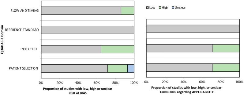
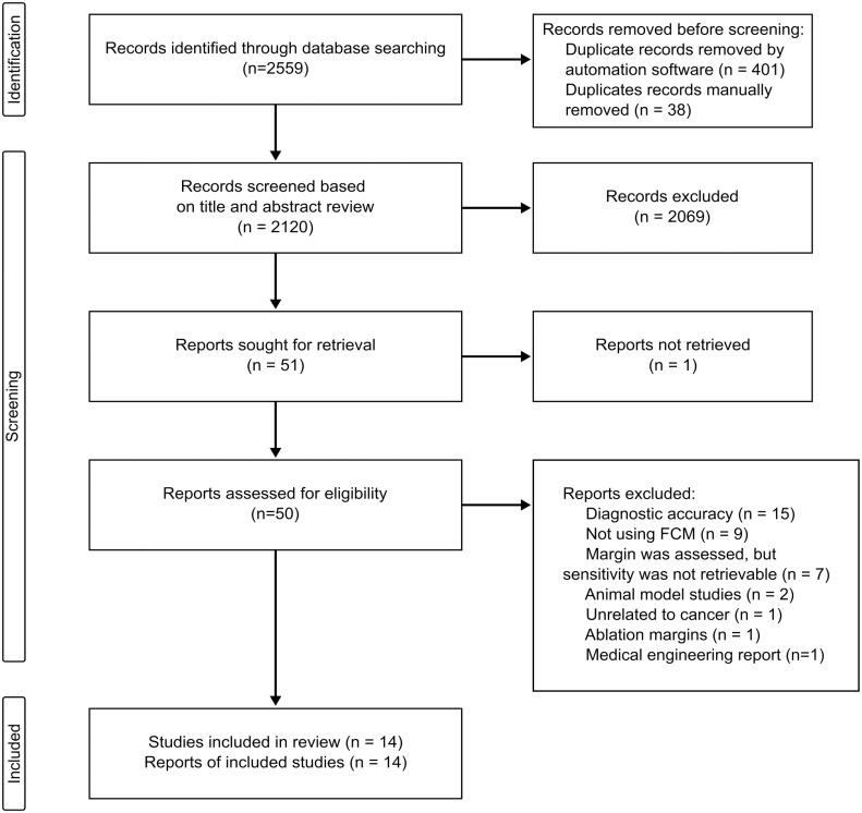
{"title":"Accuracy of Ex-vivo Fluorescence Confocal Microscopy in Margin Assessment of Solid Tumors: A Systematic Review.","authors":"Matthew Au, Ricardo Almeida-Magana, Tarek Al-Hammouri, Aiman Haider, Greg Shaw","doi":"10.1369/00221554231212948","DOIUrl":null,"url":null,"abstract":"<p><p>Fluorescence confocal microscopy (FCM) is a novel technology that enables rapid high-resolution digital imaging of non-formalin-fixed tissue specimens and offers real-time positive surgical margin identification. In this systematic review, we evaluated the accuracy metrics of ex vivo FCM for intraoperative margin assessment of different tumor types. A systematic search of MEDLINE via PubMed, Embase, Cochrane Central Register of Controlled Trials, Web of Science, and Scopus was performed for relevant papers (PROSPERO ID: CRD42022372558). We included 14 studies evaluating four types of microscopes in six different tumor types, including breast, prostate, central nervous system, kidney, bladder, and conjunctival tumors. Using the Quality Assessment of Diagnostic Accuracy Studies tool, we identified a high risk of bias in patient selection (21%) and index test (36%) of the included studies. Overall, we found that FCM has good accuracy metrics in all tumor types, with high sensitivity and specificity (>80%) and almost perfect concordance (>90%) against final pathology results. Despite these promising findings, the quality of the available evidence and bias concerns highlight the need for adequately designed studies to further define the role of ex vivo FCM in replacing the frozen section as the tool of choice for intraoperative margin assessment.</p>","PeriodicalId":16079,"journal":{"name":"Journal of Histochemistry & Cytochemistry","volume":" ","pages":"661-674"},"PeriodicalIF":1.5000,"publicationDate":"2023-12-01","publicationTypes":"Journal Article","fieldsOfStudy":null,"isOpenAccess":false,"openAccessPdf":"https://www.ncbi.nlm.nih.gov/pmc/articles/PMC10691410/pdf/","citationCount":"0","resultStr":null,"platform":"Semanticscholar","paperid":null,"PeriodicalName":"Journal of Histochemistry & Cytochemistry","FirstCategoryId":"99","ListUrlMain":"https://doi.org/10.1369/00221554231212948","RegionNum":4,"RegionCategory":"生物学","ArticlePicture":[],"TitleCN":null,"AbstractTextCN":null,"PMCID":null,"EPubDate":"2023/11/15 0:00:00","PubModel":"Epub","JCR":"Q4","JCRName":"CELL BIOLOGY","Score":null,"Total":0}
引用次数: 0
Abstract
Fluorescence confocal microscopy (FCM) is a novel technology that enables rapid high-resolution digital imaging of non-formalin-fixed tissue specimens and offers real-time positive surgical margin identification. In this systematic review, we evaluated the accuracy metrics of ex vivo FCM for intraoperative margin assessment of different tumor types. A systematic search of MEDLINE via PubMed, Embase, Cochrane Central Register of Controlled Trials, Web of Science, and Scopus was performed for relevant papers (PROSPERO ID: CRD42022372558). We included 14 studies evaluating four types of microscopes in six different tumor types, including breast, prostate, central nervous system, kidney, bladder, and conjunctival tumors. Using the Quality Assessment of Diagnostic Accuracy Studies tool, we identified a high risk of bias in patient selection (21%) and index test (36%) of the included studies. Overall, we found that FCM has good accuracy metrics in all tumor types, with high sensitivity and specificity (>80%) and almost perfect concordance (>90%) against final pathology results. Despite these promising findings, the quality of the available evidence and bias concerns highlight the need for adequately designed studies to further define the role of ex vivo FCM in replacing the frozen section as the tool of choice for intraoperative margin assessment.
荧光共聚焦显微镜(FCM)是一种新技术,可以实现非福尔马林固定组织标本的快速高分辨率数字成像,并提供实时阳性手术边缘识别。在这篇系统综述中,我们评估了体外FCM在不同肿瘤类型术中边缘评估中的准确性指标。通过PubMed、Embase、Cochrane Central Register of Controlled Trials、Web of Science和Scopus系统检索MEDLINE的相关论文(PROSPERO ID: CRD42022372558)。我们纳入了14项研究,评估了四种显微镜在六种不同肿瘤类型中的应用,包括乳腺、前列腺、中枢神经系统、肾脏、膀胱和结膜肿瘤。使用诊断准确性研究的质量评估工具,我们在纳入的研究中确定了患者选择(21%)和指数测试(36%)的高偏倚风险。总体而言,我们发现FCM在所有肿瘤类型中都具有良好的准确性指标,具有高灵敏度和特异性(>80%),与最终病理结果几乎完全一致(>90%)。尽管有这些令人鼓舞的发现,但现有证据的质量和对偏倚的担忧突出表明,需要进行充分设计的研究,以进一步确定体外FCM在替代冷冻切片作为术中边缘评估选择工具方面的作用。
期刊介绍:
Journal of Histochemistry & Cytochemistry (JHC) has been a pre-eminent cell biology journal for over 50 years. Published monthly, JHC offers primary research articles, timely reviews, editorials, and perspectives on the structure and function of cells, tissues, and organs, as well as mechanisms of development, differentiation, and disease. JHC also publishes new developments in microscopy and imaging, especially where imaging techniques complement current genetic, molecular and biochemical investigations of cell and tissue function. JHC offers generous space for articles and recognizing the value of images that reveal molecular, cellular and tissue organization, offers free color to all authors.


 求助内容:
求助内容: 应助结果提醒方式:


