Ren Jie Jacob Chew, Jacinta Xiaotong Lu, Yu Fan Sim, Alvin Boon Keng Yeo
{"title":"Rodent peri-implantitis models: a systematic review and meta-analysis of morphological changes.","authors":"Ren Jie Jacob Chew, Jacinta Xiaotong Lu, Yu Fan Sim, Alvin Boon Keng Yeo","doi":"10.5051/jpis.2200900045","DOIUrl":null,"url":null,"abstract":"<p><strong>Purpose: </strong>Rodent models have emerged as an alternative to established larger animal models for peri-implantitis research. However, the construct validity of rodent models is controversial due to a lack of consensus regarding their histological, morphological, and biochemical characteristics. This systematic review sought to validate rodent models by characterizing their morphological changes, particularly marginal bone loss (MBL), a hallmark of peri-implantitis.</p><p><strong>Methods: </strong>This review was conducted following the Preferred Reporting Items for Systematic Reviews and Meta-Analysis (PRISMA) guidelines. A literature search was performed electronically using MEDLINE (PubMed), and Embase, identifying pre-clinical studies reporting MBL after experimental peri-implantitis induction in rodents. Each study's risk of bias was assessed using the Systematic Review Center for Laboratory animal Experimentation (SYRCLE) risk of bias tool. A meta-analysis was performed for the difference in MBL, comparing healthy implants to those with experimental peri-implantitis.</p><p><strong>Results: </strong>Of the 1,014 unique records retrieved, 23 studies that met the eligibility criteria were included. Peri-implantitis was induced using 4 methods: ligatures, lipopolysaccharide, microbial infection, and titanium particles. Studies presented high to unclear risks of bias. During the osseointegration phase, 11.6% and 6.4%-11.3% of implants inserted in mice and rats, respectively, had failed to osseointegrate. Twelve studies were included in the meta-analysis of the linear MBL measured using micro-computed tomography. Following experimental peri-implantitis, the MBL was estimated to be 0.25 mm (95% confidence interval [CI], 0.14-0.36 mm) in mice and 0.26 mm (95% CI, 0.19-0.34 mm) in rats. The resulting peri-implant MBL was circumferential, consisting of supra- and infrabony components.</p><p><strong>Conclusions: </strong>Experimental peri-implantitis in rodent models results in circumferential MBL, with morphology consistent with the clinical presentation of peri-implantitis. While rodent models are promising, there is still a need to further characterize their healing potentials, standardize experiment protocols, and improve the reporting of results and methodology.</p><p><strong>Trial registration: </strong>PROSPERO Identifier: CRD42020209776.</p>","PeriodicalId":48795,"journal":{"name":"Journal of Periodontal and Implant Science","volume":"52 6","pages":"479-495"},"PeriodicalIF":2.2000,"publicationDate":"2022-12-01","publicationTypes":"Journal Article","fieldsOfStudy":null,"isOpenAccess":false,"openAccessPdf":"https://ftp.ncbi.nlm.nih.gov/pub/pmc/oa_pdf/d9/ae/jpis-52-479.PMC9807853.pdf","citationCount":"1","resultStr":null,"platform":"Semanticscholar","paperid":null,"PeriodicalName":"Journal of Periodontal and Implant Science","FirstCategoryId":"3","ListUrlMain":"https://doi.org/10.5051/jpis.2200900045","RegionNum":4,"RegionCategory":"医学","ArticlePicture":[],"TitleCN":null,"AbstractTextCN":null,"PMCID":null,"EPubDate":"","PubModel":"","JCR":"Q2","JCRName":"DENTISTRY, ORAL SURGERY & MEDICINE","Score":null,"Total":0}
引用次数: 1
Abstract
Purpose: Rodent models have emerged as an alternative to established larger animal models for peri-implantitis research. However, the construct validity of rodent models is controversial due to a lack of consensus regarding their histological, morphological, and biochemical characteristics. This systematic review sought to validate rodent models by characterizing their morphological changes, particularly marginal bone loss (MBL), a hallmark of peri-implantitis.
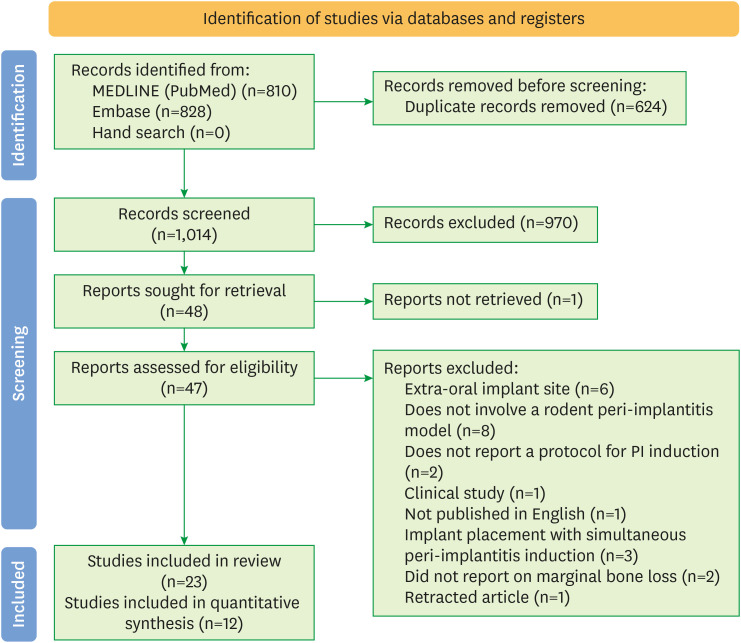
Methods: This review was conducted following the Preferred Reporting Items for Systematic Reviews and Meta-Analysis (PRISMA) guidelines. A literature search was performed electronically using MEDLINE (PubMed), and Embase, identifying pre-clinical studies reporting MBL after experimental peri-implantitis induction in rodents. Each study's risk of bias was assessed using the Systematic Review Center for Laboratory animal Experimentation (SYRCLE) risk of bias tool. A meta-analysis was performed for the difference in MBL, comparing healthy implants to those with experimental peri-implantitis.
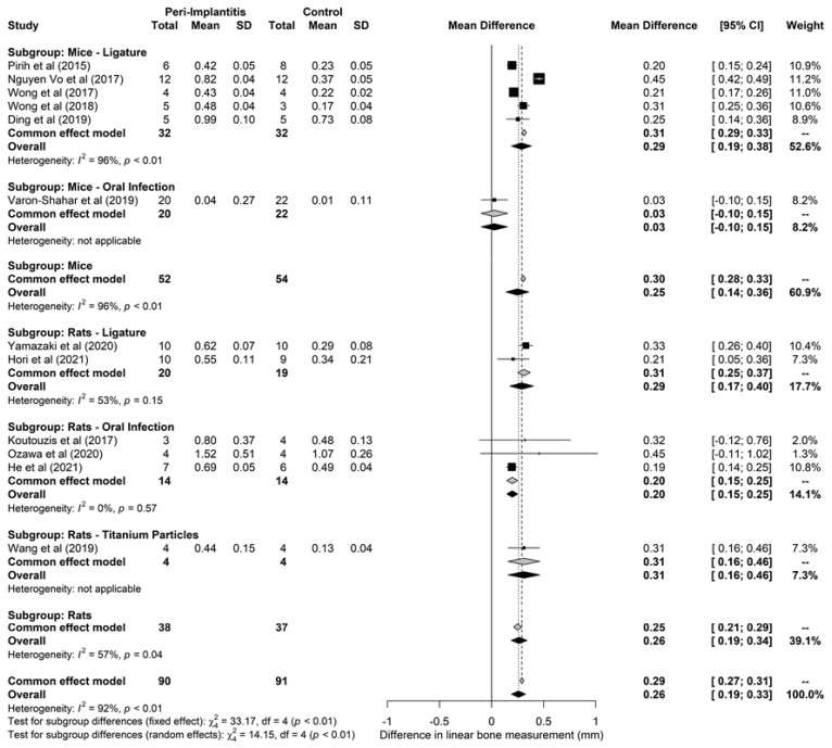
Results: Of the 1,014 unique records retrieved, 23 studies that met the eligibility criteria were included. Peri-implantitis was induced using 4 methods: ligatures, lipopolysaccharide, microbial infection, and titanium particles. Studies presented high to unclear risks of bias. During the osseointegration phase, 11.6% and 6.4%-11.3% of implants inserted in mice and rats, respectively, had failed to osseointegrate. Twelve studies were included in the meta-analysis of the linear MBL measured using micro-computed tomography. Following experimental peri-implantitis, the MBL was estimated to be 0.25 mm (95% confidence interval [CI], 0.14-0.36 mm) in mice and 0.26 mm (95% CI, 0.19-0.34 mm) in rats. The resulting peri-implant MBL was circumferential, consisting of supra- and infrabony components.
Conclusions: Experimental peri-implantitis in rodent models results in circumferential MBL, with morphology consistent with the clinical presentation of peri-implantitis. While rodent models are promising, there is still a need to further characterize their healing potentials, standardize experiment protocols, and improve the reporting of results and methodology.
期刊介绍:
Journal of Periodontal & Implant Science (JPIS) is a peer-reviewed and open-access journal providing up-to-date information relevant to professionalism of periodontology and dental implantology. JPIS is dedicated to global and extensive publication which includes evidence-based original articles, and fundamental reviews in order to cover a variety of interests in the field of periodontal as well as implant science.

 求助内容:
求助内容: 应助结果提醒方式:


