Erfan Saatchian, Sina Ehsani, Alireza Montazerabadi
{"title":"Evaluation of Two Post-Processing Analysis Methods of Proton Magnetic Resonance Spectroscopy in Glioma Tumors.","authors":"Erfan Saatchian, Sina Ehsani, Alireza Montazerabadi","doi":"10.31661/jbpe.v0i0.2001-1055","DOIUrl":null,"url":null,"abstract":"<p><strong>Background: </strong>Magnetic resonance spectroscopy (MRS) is a non-invasive diagnostic and the neuroimaging method of choice for the noninvasive monitoring of brain metabolism in patients with glioma tumors. <sup>1</sup>H-MRS is a reliable and non-invasive tool used to study glioma. However, the metabolite spectra obtained by <sup>1</sup>H-MRS requires a specific quantification procedure for post-processing. According to our knowledge, no comparisons have yet been made between spectrum analysis software for quantification of gliomas metabolites.</p><p><strong>Objective: </strong>Current study aims to evaluate the difference between this two common software in quantifying cerebral metabolites.</p><p><strong>Material and methods: </strong>In this analytical study, we evaluate two post-processing software packages, java-based graphical for MR user interface packages (jMRUI) and totally automatic robust quantitation in NMR (TARQUIN) software. <sup>1</sup>H-MRS spectrum from the brain of patients with gliomas tumors was collected for post-processing. AMARES algorithms were conducted to metabolite qualification on jMRUI software, and TARQUIN software were implemented with automated quantification algorithms. The study included a total of 30 subjects. For quantification, subjects were divided into a normal group (n=15) and group of gliomas (n=15).</p><p><strong>Results: </strong>When calculated by TARQUIN, the mean metabolites ratio was typically lower than by jMRUI. While, the mean ratio of metabolites varied when quantified by jMRUI vs. TARQUIN, both methods apparent clinical associations.</p><p><strong>Conclusion: </strong>TARQUIN and jMRUI are feasible choices for the post-processing of cerebral MRS data obtained from glioma tumors.</p>","PeriodicalId":38035,"journal":{"name":"Journal of Biomedical Physics and Engineering","volume":null,"pages":null},"PeriodicalIF":0.0000,"publicationDate":"2023-02-01","publicationTypes":"Journal Article","fieldsOfStudy":null,"isOpenAccess":false,"openAccessPdf":"https://ftp.ncbi.nlm.nih.gov/pub/pmc/oa_pdf/c4/b2/JBPE-13-39.PMC9923243.pdf","citationCount":"1","resultStr":null,"platform":"Semanticscholar","paperid":null,"PeriodicalName":"Journal of Biomedical Physics and Engineering","FirstCategoryId":"1085","ListUrlMain":"https://doi.org/10.31661/jbpe.v0i0.2001-1055","RegionNum":0,"RegionCategory":null,"ArticlePicture":[],"TitleCN":null,"AbstractTextCN":null,"PMCID":null,"EPubDate":"","PubModel":"","JCR":"Q3","JCRName":"Medicine","Score":null,"Total":0}
引用次数: 1
Abstract
Background: Magnetic resonance spectroscopy (MRS) is a non-invasive diagnostic and the neuroimaging method of choice for the noninvasive monitoring of brain metabolism in patients with glioma tumors. 1H-MRS is a reliable and non-invasive tool used to study glioma. However, the metabolite spectra obtained by 1H-MRS requires a specific quantification procedure for post-processing. According to our knowledge, no comparisons have yet been made between spectrum analysis software for quantification of gliomas metabolites.
Objective: Current study aims to evaluate the difference between this two common software in quantifying cerebral metabolites.
Material and methods: In this analytical study, we evaluate two post-processing software packages, java-based graphical for MR user interface packages (jMRUI) and totally automatic robust quantitation in NMR (TARQUIN) software. 1H-MRS spectrum from the brain of patients with gliomas tumors was collected for post-processing. AMARES algorithms were conducted to metabolite qualification on jMRUI software, and TARQUIN software were implemented with automated quantification algorithms. The study included a total of 30 subjects. For quantification, subjects were divided into a normal group (n=15) and group of gliomas (n=15).
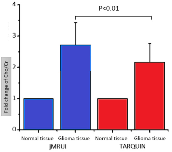
Results: When calculated by TARQUIN, the mean metabolites ratio was typically lower than by jMRUI. While, the mean ratio of metabolites varied when quantified by jMRUI vs. TARQUIN, both methods apparent clinical associations.
Conclusion: TARQUIN and jMRUI are feasible choices for the post-processing of cerebral MRS data obtained from glioma tumors.
期刊介绍:
The Journal of Biomedical Physics and Engineering (JBPE) is a bimonthly peer-reviewed English-language journal that publishes high-quality basic sciences and clinical research (experimental or theoretical) broadly concerned with the relationship of physics to medicine and engineering.


 求助内容:
求助内容: 应助结果提醒方式:


