Matthijs Snelders , Iris H. Koedijk , Julia Schirmer , Otto Mulleners , Juancito van Leeuwen , Nathalie P. de Wagenaar , Oscar Bartulos , Pieter Voskamp , Stefan Braam , Zeno Guttenberg , A.H. Jan Danser , Danielle Majoor-Krakauer , Erik Meijering , Ingrid van der Pluijm , Jeroen Essers
{"title":"Contraction pressure analysis using optical imaging in normal and MYBPC3-mutated hiPSC-derived cardiomyocytes grown on matrices with tunable stiffness","authors":"Matthijs Snelders , Iris H. Koedijk , Julia Schirmer , Otto Mulleners , Juancito van Leeuwen , Nathalie P. de Wagenaar , Oscar Bartulos , Pieter Voskamp , Stefan Braam , Zeno Guttenberg , A.H. Jan Danser , Danielle Majoor-Krakauer , Erik Meijering , Ingrid van der Pluijm , Jeroen Essers","doi":"10.1016/j.bbiosy.2022.100068","DOIUrl":null,"url":null,"abstract":"<div><p>Current <em>in vivo</em> disease models and analysis methods for cardiac drug development have been insufficient in providing accurate and reliable predictions of drug efficacy and safety. Here, we propose a custom optical flow-based analysis method to quantitatively measure recordings of contracting cardiomyocytes on polydimethylsiloxane (PDMS), compatible with medium-throughput systems.</p><p>Movement of the PDMS was examined by covalently bound fluorescent beads on the PDMS surface, differences caused by increased substrate stiffness were compared, and cells were stimulated with β-agonist. We further validated the system using cardiomyocytes treated with endothelin-1 and compared their contractions against control and cells incubated with receptor antagonist bosentan. After validation we examined two MYBPC3-mutant patient-derived cell lines.</p><p>Recordings showed that higher substrate stiffness resulted in higher contractile pressure, while beating frequency remained similar to control. β-agonist stimulation resulted in both higher beating frequency as well as higher pressure values during contraction and relaxation. Cells treated with endothelin-1 showed an increased beating frequency, but a lower contraction pressure. Cells treated with both endothelin-1 and bosentan remained at control level of beating frequency and pressure. Lastly, both MYBPC3-mutant lines showed a higher beating frequency and lower contraction pressure.</p><p>Our validated method is capable of automatically quantifying contraction of hiPSC-derived cardiomyocytes on a PDMS substrate of known shear modulus, returning an absolute value. Our method could have major benefits in a medium-throughput setting.</p></div>","PeriodicalId":72379,"journal":{"name":"Biomaterials and biosystems","volume":"8 ","pages":"Article 100068"},"PeriodicalIF":0.0000,"publicationDate":"2022-12-01","publicationTypes":"Journal Article","fieldsOfStudy":null,"isOpenAccess":false,"openAccessPdf":"https://ftp.ncbi.nlm.nih.gov/pub/pmc/oa_pdf/3a/2b/main.PMC9934435.pdf","citationCount":"1","resultStr":null,"platform":"Semanticscholar","paperid":null,"PeriodicalName":"Biomaterials and biosystems","FirstCategoryId":"1085","ListUrlMain":"https://www.sciencedirect.com/science/article/pii/S2666534422000307","RegionNum":0,"RegionCategory":null,"ArticlePicture":[],"TitleCN":null,"AbstractTextCN":null,"PMCID":null,"EPubDate":"","PubModel":"","JCR":"Q3","JCRName":"Biochemistry, Genetics and Molecular Biology","Score":null,"Total":0}
引用次数: 1
Abstract
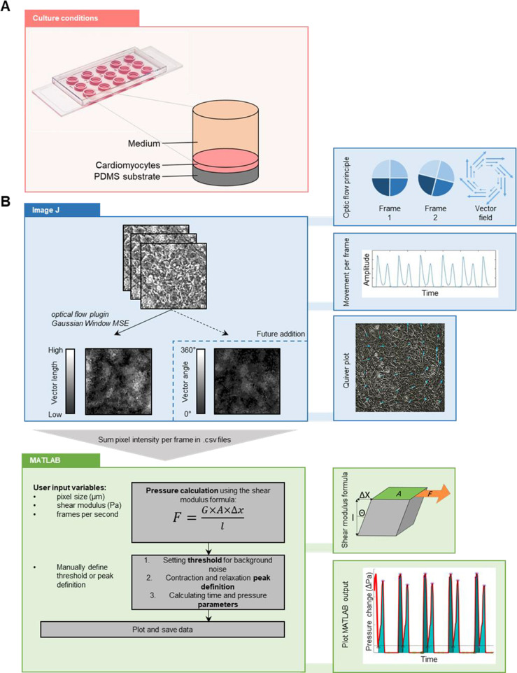
Current in vivo disease models and analysis methods for cardiac drug development have been insufficient in providing accurate and reliable predictions of drug efficacy and safety. Here, we propose a custom optical flow-based analysis method to quantitatively measure recordings of contracting cardiomyocytes on polydimethylsiloxane (PDMS), compatible with medium-throughput systems.
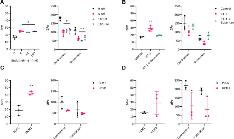
Movement of the PDMS was examined by covalently bound fluorescent beads on the PDMS surface, differences caused by increased substrate stiffness were compared, and cells were stimulated with β-agonist. We further validated the system using cardiomyocytes treated with endothelin-1 and compared their contractions against control and cells incubated with receptor antagonist bosentan. After validation we examined two MYBPC3-mutant patient-derived cell lines.
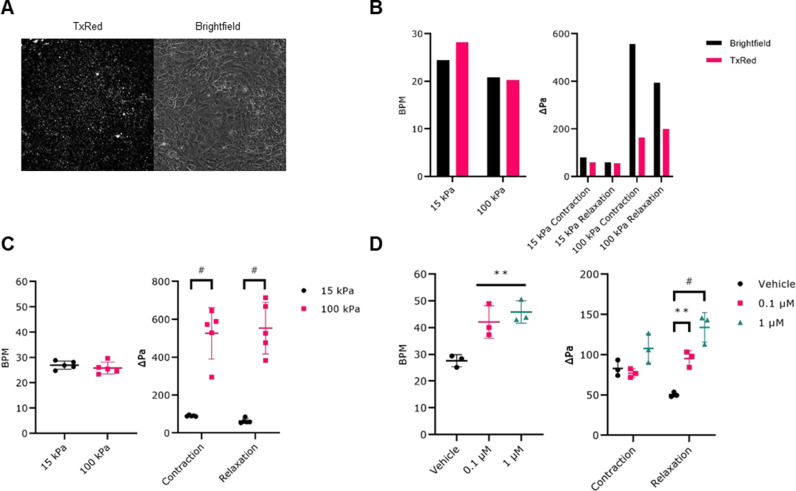
Recordings showed that higher substrate stiffness resulted in higher contractile pressure, while beating frequency remained similar to control. β-agonist stimulation resulted in both higher beating frequency as well as higher pressure values during contraction and relaxation. Cells treated with endothelin-1 showed an increased beating frequency, but a lower contraction pressure. Cells treated with both endothelin-1 and bosentan remained at control level of beating frequency and pressure. Lastly, both MYBPC3-mutant lines showed a higher beating frequency and lower contraction pressure.
Our validated method is capable of automatically quantifying contraction of hiPSC-derived cardiomyocytes on a PDMS substrate of known shear modulus, returning an absolute value. Our method could have major benefits in a medium-throughput setting.

 求助内容:
求助内容: 应助结果提醒方式:


