Ilona Mihaela Liliac, Mihai Victor Sacerdoțianu, Bogdan Silviu Ungureanu, Laurențiu Mogoantă, Claudiu Mărgăritescu, Daniel Pirici, Adrian Săftoiu, Anda Elena Crisan
{"title":"Subtle Immunoreactivity Differences in the Fractal Patterns of Membrane E-Cadherin in Gastric Adenocarcinoma.","authors":"Ilona Mihaela Liliac, Mihai Victor Sacerdoțianu, Bogdan Silviu Ungureanu, Laurențiu Mogoantă, Claudiu Mărgăritescu, Daniel Pirici, Adrian Săftoiu, Anda Elena Crisan","doi":"10.12865/CHSJ.48.03.08","DOIUrl":null,"url":null,"abstract":"<p><p>Gastric cancer continues to be a significant malignancy worldwide, accounting for approximately one million new cases in 2020. Scientists are focusing on the cancerous cells' plasma membrane (PM) as a potential therapeutic target in cancer because it functions as the cell's interface with its environment through a variety of mechanisms. The capacity of membrane shape and its structures to influence biological processes frequently occurs through the regulation of enzymes or preferential protein binding to membranes via membrane shape changes. We aimed here to assess the morphological irregularities of the cellular membranes in gastric adenocarcinoma tumors, and to find any putative differences from normal gastric mucosae epithelial cells. We analyzed the pattern of E-cadherin at the level of the cell membrane using the fractal dimension (FD) analysis on fluorescence immunohistochemistry samples labeled with E-cadherin in gastric well/moderate and solid gastric adenocarcinoma from patients without any associated chemotherapeutic treatment or radiotherapy. Images were binarized based on a fixed threshold of the E-cadherin fluorescence channel, and then the FD of the binarized image outlines has been calculated in order to assess the ruggedness of the cellular membranes. Overall assessment of the FD revealed that the subtle membrane variations were evident enough to deem a statistically significant difference and the complexity of the membrane roughness was clearly higher for adenocarcinoma cases. We intended to evaluate if separating adenocarcinoma cases as low grade (G1 and G2) and high grade (G3 and solid), FD analysis could still differentiate membrane patterns and check if the available clinical parameters like age, gender, tumor location, lymph ganglia involved might correlate with FD values for adenocarcinoma patients. Altogether, the morphological analysis of a simple marker for the cell membrane can identify and distinguish tumor cells. Although there was a limited correlation between this analysis and the main clinical and pathological indicators of the disease, it will be very useful in the future for automatic computer-assisted diagnosis on slides, as well as for evaluating cellular adhesion and inter-cellular trafficking in cancer cells.</p>","PeriodicalId":10938,"journal":{"name":"Current Health Sciences Journal","volume":"48 3","pages":"303-310"},"PeriodicalIF":0.0000,"publicationDate":"2022-07-01","publicationTypes":"Journal Article","fieldsOfStudy":null,"isOpenAccess":false,"openAccessPdf":"https://ftp.ncbi.nlm.nih.gov/pub/pmc/oa_pdf/bc/fa/CHSJ-48-03-303.PMC9940928.pdf","citationCount":"2","resultStr":null,"platform":"Semanticscholar","paperid":null,"PeriodicalName":"Current Health Sciences Journal","FirstCategoryId":"1085","ListUrlMain":"https://doi.org/10.12865/CHSJ.48.03.08","RegionNum":0,"RegionCategory":null,"ArticlePicture":[],"TitleCN":null,"AbstractTextCN":null,"PMCID":null,"EPubDate":"2022/9/30 0:00:00","PubModel":"Epub","JCR":"","JCRName":"","Score":null,"Total":0}
引用次数: 2
Abstract
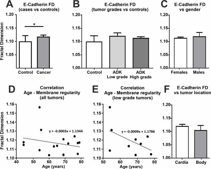
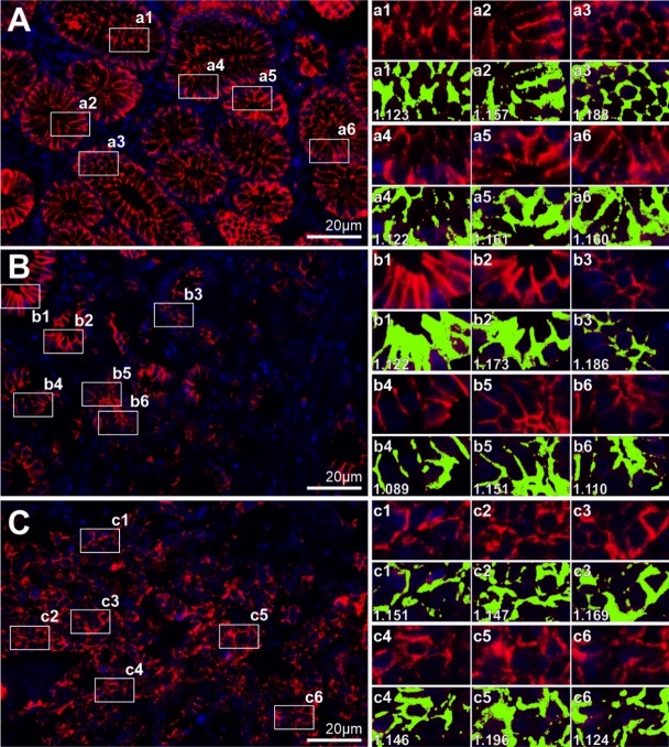
Gastric cancer continues to be a significant malignancy worldwide, accounting for approximately one million new cases in 2020. Scientists are focusing on the cancerous cells' plasma membrane (PM) as a potential therapeutic target in cancer because it functions as the cell's interface with its environment through a variety of mechanisms. The capacity of membrane shape and its structures to influence biological processes frequently occurs through the regulation of enzymes or preferential protein binding to membranes via membrane shape changes. We aimed here to assess the morphological irregularities of the cellular membranes in gastric adenocarcinoma tumors, and to find any putative differences from normal gastric mucosae epithelial cells. We analyzed the pattern of E-cadherin at the level of the cell membrane using the fractal dimension (FD) analysis on fluorescence immunohistochemistry samples labeled with E-cadherin in gastric well/moderate and solid gastric adenocarcinoma from patients without any associated chemotherapeutic treatment or radiotherapy. Images were binarized based on a fixed threshold of the E-cadherin fluorescence channel, and then the FD of the binarized image outlines has been calculated in order to assess the ruggedness of the cellular membranes. Overall assessment of the FD revealed that the subtle membrane variations were evident enough to deem a statistically significant difference and the complexity of the membrane roughness was clearly higher for adenocarcinoma cases. We intended to evaluate if separating adenocarcinoma cases as low grade (G1 and G2) and high grade (G3 and solid), FD analysis could still differentiate membrane patterns and check if the available clinical parameters like age, gender, tumor location, lymph ganglia involved might correlate with FD values for adenocarcinoma patients. Altogether, the morphological analysis of a simple marker for the cell membrane can identify and distinguish tumor cells. Although there was a limited correlation between this analysis and the main clinical and pathological indicators of the disease, it will be very useful in the future for automatic computer-assisted diagnosis on slides, as well as for evaluating cellular adhesion and inter-cellular trafficking in cancer cells.

 求助内容:
求助内容: 应助结果提醒方式:


