Stimulation of distinct parietal locations differentiates frontal versus hippocampal network involvement in memory formation

Shruti Dave , Stephen VanHaerents , Borna Bonakdarpour , M.- Marsel Mesulam , Joel L. Voss
{"title":"Stimulation of distinct parietal locations differentiates frontal versus hippocampal network involvement in memory formation","authors":"Shruti Dave ,&nbsp;Stephen VanHaerents ,&nbsp;Borna Bonakdarpour ,&nbsp;M.- Marsel Mesulam ,&nbsp;Joel L. Voss","doi":"10.1016/j.crneur.2022.100030","DOIUrl":null,"url":null,"abstract":"<div><p>Adjacent regions of parietal cortex are thought to affiliate with distinct large-scale networks and thereby make different contributions to memory formation. We directly tested this putative functional segregation within parietal cortex by perturbing activity of anterior versus posterior parietal areas. We applied noninvasive theta-burst transcranial magnetic stimulation to these locations immediately before a semantic encoding task, and subsequently tested recollection memory. Consistent with previous findings, fMRI activity in left inferior frontal gyrus during semantic encoding correlated with subsequent high memory accuracy and strong subjective recollection. Stimulation of the posterior parietal cortex decoupled its network – the hippocampal-cortical network – from left inferior frontal gyrus. Furthermore, posterior parietal stimulation reduced highly accurate subjective recollection. Critically, both of these changes occurred relative to stimulation of the anterior parietal cortex. Stimulating anterior versus posterior parietal cortex therefore differentiated hippocampal network involvement in episodic memory. This provides direct evidence that distinct territories within close proximity of each other in parietal cortex make functionally distinct contributions to memory formation. Further, noninvasive stimulation has the spatial resolution required to differentially modulate the interaction of these adjacent parietal locations with distributed large-scale brain networks.</p></div>","PeriodicalId":72752,"journal":{"name":"Current research in neurobiology","volume":"3 ","pages":"Article 100030"},"PeriodicalIF":0.0000,"publicationDate":"2022-01-01","publicationTypes":"Journal Article","fieldsOfStudy":null,"isOpenAccess":false,"openAccessPdf":"https://ftp.ncbi.nlm.nih.gov/pub/pmc/oa_pdf/3e/b0/main.PMC9743066.pdf","citationCount":"0","resultStr":null,"platform":"Semanticscholar","paperid":null,"PeriodicalName":"Current research in neurobiology","FirstCategoryId":"1085","ListUrlMain":"https://www.sciencedirect.com/science/article/pii/S2665945X22000031","RegionNum":0,"RegionCategory":null,"ArticlePicture":[],"TitleCN":null,"AbstractTextCN":null,"PMCID":null,"EPubDate":"","PubModel":"","JCR":"","JCRName":"","Score":null,"Total":0}
引用次数: 0

Abstract

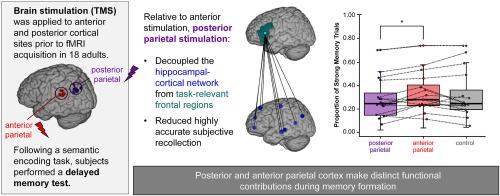
Adjacent regions of parietal cortex are thought to affiliate with distinct large-scale networks and thereby make different contributions to memory formation. We directly tested this putative functional segregation within parietal cortex by perturbing activity of anterior versus posterior parietal areas. We applied noninvasive theta-burst transcranial magnetic stimulation to these locations immediately before a semantic encoding task, and subsequently tested recollection memory. Consistent with previous findings, fMRI activity in left inferior frontal gyrus during semantic encoding correlated with subsequent high memory accuracy and strong subjective recollection. Stimulation of the posterior parietal cortex decoupled its network – the hippocampal-cortical network – from left inferior frontal gyrus. Furthermore, posterior parietal stimulation reduced highly accurate subjective recollection. Critically, both of these changes occurred relative to stimulation of the anterior parietal cortex. Stimulating anterior versus posterior parietal cortex therefore differentiated hippocampal network involvement in episodic memory. This provides direct evidence that distinct territories within close proximity of each other in parietal cortex make functionally distinct contributions to memory formation. Further, noninvasive stimulation has the spatial resolution required to differentially modulate the interaction of these adjacent parietal locations with distributed large-scale brain networks.
Abstract Image

不同顶叶位置的刺激区分额叶与海马网络参与记忆形成
顶叶皮层的邻近区域被认为与不同的大规模网络相关联,因此对记忆的形成有不同的贡献。我们通过干扰前顶叶区和后顶叶区的活动,直接测试了顶叶皮层中这种假定的功能分离。在语义编码任务之前,我们对这些部位进行了非侵入性的经颅磁刺激,随后测试了回忆记忆。与先前的研究结果一致,语义编码过程中左侧额下回的fMRI活动与随后的高记忆准确性和强主观回忆相关。后顶叶皮层的刺激使其网络——海马体-皮质网络——与左侧额下回分离。此外,后顶叶刺激降低了高度准确的主观回忆。关键的是,这两种变化都与刺激前顶叶皮层有关。刺激前顶叶和后顶叶皮层因此区分海马网络参与情景记忆。这提供了直接证据,表明顶叶皮层中彼此接近的不同区域在功能上对记忆的形成有不同的贡献。此外,非侵入性刺激具有空间分辨率,可以不同地调节这些相邻顶叶位置与分布式大尺度脑网络之间的相互作用。
本文章由计算机程序翻译,如有差异,请以英文原文为准。
求助全文
约1分钟内获得全文 求助全文
来源期刊
CiteScore
2.20
自引率
0.00%
发文量
0
×
引用
GB/T 7714-2015
复制
MLA
复制
APA
复制
导出至
BibTeX EndNote RefMan NoteFirst NoteExpress
×
提示
您的信息不完整,为了账户安全,请先补充。
现在去补充
×
提示
您因"违规操作"
具体请查看互助需知
我知道了
×
提示
确定
请完成安全验证×
copy
已复制链接
快去分享给好友吧!
我知道了
右上角分享
点击右上角分享
0
联系我们:info@booksci.cn Book学术提供免费学术资源搜索服务,方便国内外学者检索中英文文献。致力于提供最便捷和优质的服务体验。 Copyright © 2023 布克学术 All rights reserved.
京ICP备2023020795号-1
ghs 京公网安备 11010802042870号
Book学术文献互助
Book学术文献互助群
群 号:481959085
Book学术官方微信