Dmitrij Kvitka, Dalia Juodžentė, Greta Rudenkovaitė, Evelina Burbaitė, Monika Laukutė
{"title":"Successful early diagnosis and surgical treatment of congenital caval foramen hernia in an 8-month-old mixed breed cat.","authors":"Dmitrij Kvitka, Dalia Juodžentė, Greta Rudenkovaitė, Evelina Burbaitė, Monika Laukutė","doi":"10.29374/2527-2179.bjvm005622","DOIUrl":null,"url":null,"abstract":"<p><p>An 8-month-old neutered female domestic mixed breed cat was presented to Dr. L. Kriaučeliūnas Small Animal Clinic due to coughing that persisted for 2 weeks. Lateral and dorsoventral chest radiographs revealed an unusual dome-shaped soft tissue opacity mass that had contact with the cranial part of the diaphragm. Together with heart and abdominal ultrasound findings, we decided that one of the differential diagnoses was a diaphragmatic hernia. During the diagnostic celiotomy, a vertical 4 cm in length diaphragmatic deficit was visualized. Left medial and lateral liver lobes were herniated, yet healthy-looking. Adhesions between the liver lobes and the pericardium sac were visualized and dissected. The pericardium was sutured with simple interrupted suture pattern. A herniorrhaphy was performed suturing the diaphragm with the continuous suture pattern. Successful surgical treatment resulted in fully resolved clinical symptoms.</p>","PeriodicalId":72458,"journal":{"name":"Brazilian journal of veterinary medicine","volume":"45 ","pages":"e005622"},"PeriodicalIF":0.0000,"publicationDate":"2023-01-01","publicationTypes":"Journal Article","fieldsOfStudy":null,"isOpenAccess":false,"openAccessPdf":"https://ftp.ncbi.nlm.nih.gov/pub/pmc/oa_pdf/db/9a/bjvm-45-e005622.PMC9910220.pdf","citationCount":"0","resultStr":null,"platform":"Semanticscholar","paperid":null,"PeriodicalName":"Brazilian journal of veterinary medicine","FirstCategoryId":"1085","ListUrlMain":"https://doi.org/10.29374/2527-2179.bjvm005622","RegionNum":0,"RegionCategory":null,"ArticlePicture":[],"TitleCN":null,"AbstractTextCN":null,"PMCID":null,"EPubDate":"","PubModel":"","JCR":"","JCRName":"","Score":null,"Total":0}
引用次数: 0
Abstract
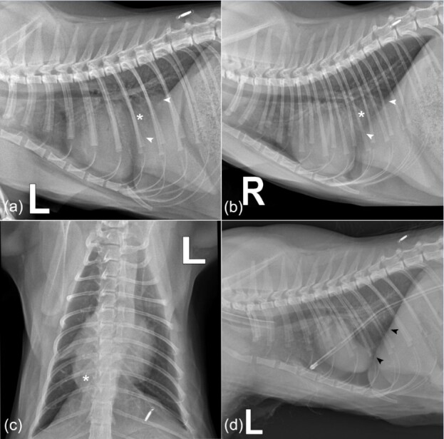
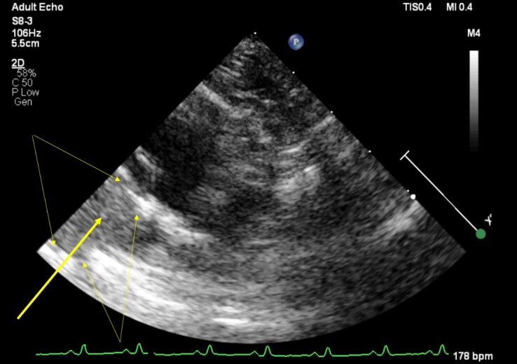
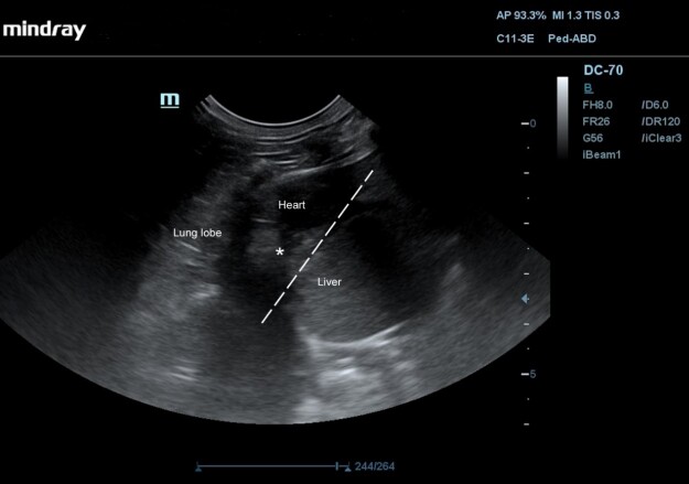
An 8-month-old neutered female domestic mixed breed cat was presented to Dr. L. Kriaučeliūnas Small Animal Clinic due to coughing that persisted for 2 weeks. Lateral and dorsoventral chest radiographs revealed an unusual dome-shaped soft tissue opacity mass that had contact with the cranial part of the diaphragm. Together with heart and abdominal ultrasound findings, we decided that one of the differential diagnoses was a diaphragmatic hernia. During the diagnostic celiotomy, a vertical 4 cm in length diaphragmatic deficit was visualized. Left medial and lateral liver lobes were herniated, yet healthy-looking. Adhesions between the liver lobes and the pericardium sac were visualized and dissected. The pericardium was sutured with simple interrupted suture pattern. A herniorrhaphy was performed suturing the diaphragm with the continuous suture pattern. Successful surgical treatment resulted in fully resolved clinical symptoms.

 求助内容:
求助内容: 应助结果提醒方式:


