Diogo G Corrêa, Eelco van Duinkerken, João Gabriel D Farinhas, Valéria C Pereira, Emerson L Gasparetto, Soniza V Alves-Leon, Fernanda Cristina R Lopes
{"title":"Influence of natalizumab on resting-state connectivity in patients with multiple sclerosis.","authors":"Diogo G Corrêa, Eelco van Duinkerken, João Gabriel D Farinhas, Valéria C Pereira, Emerson L Gasparetto, Soniza V Alves-Leon, Fernanda Cristina R Lopes","doi":"10.1177/11795735231195775","DOIUrl":null,"url":null,"abstract":"<p><strong>Background: </strong>Changes in brain connectivity occur in patients with multiple sclerosis (MS), even in patients under disease-modifying therapies. Using magnetic resonance imaging (MRI) to asses patients treated with disease-modifying therapies, such as natalizumab, can elucidate the mechanisms involved in clinical deterioration in MS.</p><p><strong>Objectives: </strong>To evaluate differences in resting-state functional connectivity among MS patients treated with natalizumab, MS patients not treated with natalizumab, and controls.</p><p><strong>Design: </strong>Single-center retrospective cross-sectional study.</p><p><strong>Methods: </strong>Twenty-three MS patients being treated with natalizumab were retrospectively compared with 23 MS patients who were naïve for natalizumab, and were using first-line medications (interferon-β and/or glatiramer acetate), and 17 gender- and age-matched control subjects. The MS patient groups were also matched for time since diagnosis and hyperintense lesion volume on FLAIR. All participants underwent brain MRI using a 3 Tesla scanner. Independent component analysis and dual regression were used to identify resting-state functional connectivity using the FMRIB Software Library.</p><p><strong>Results: </strong>In comparison to controls, the MS patients treated with natalizumab presented decreased connectivity in the left orbitofrontal cortex, in the anterior cingulate and orbitofrontal cortex network. The patients not treated with natalizumab presented increased connectivity in the secondary visual, sensorimotor, and ventral attention networks in comparison to controls.Compared to patients treated with natalizumab, the patients not using natalizumab presented increased connectivity in the left Heschl's gyrus and in the right superior frontal gyrus in the ventral attention network.</p><p><strong>Conclusion: </strong>Differences in brain connectivity between MS patients not treated with natalizumab, healthy controls, and patients treated with natalizumab may be secondary to suboptimal neuronal compensation due to prior less efficient treatments, or due to a compensation in response to maladaptive plasticity.</p>","PeriodicalId":15218,"journal":{"name":"Journal of Central Nervous System Disease","volume":"15 ","pages":"11795735231195775"},"PeriodicalIF":2.8000,"publicationDate":"2023-01-01","publicationTypes":"Journal Article","fieldsOfStudy":null,"isOpenAccess":false,"openAccessPdf":"https://ftp.ncbi.nlm.nih.gov/pub/pmc/oa_pdf/44/f0/10.1177_11795735231195775.PMC10433731.pdf","citationCount":"0","resultStr":null,"platform":"Semanticscholar","paperid":null,"PeriodicalName":"Journal of Central Nervous System Disease","FirstCategoryId":"1085","ListUrlMain":"https://doi.org/10.1177/11795735231195775","RegionNum":0,"RegionCategory":null,"ArticlePicture":[],"TitleCN":null,"AbstractTextCN":null,"PMCID":null,"EPubDate":"","PubModel":"","JCR":"Q2","JCRName":"CLINICAL NEUROLOGY","Score":null,"Total":0}
引用次数: 0
Abstract
Background: Changes in brain connectivity occur in patients with multiple sclerosis (MS), even in patients under disease-modifying therapies. Using magnetic resonance imaging (MRI) to asses patients treated with disease-modifying therapies, such as natalizumab, can elucidate the mechanisms involved in clinical deterioration in MS.
Objectives: To evaluate differences in resting-state functional connectivity among MS patients treated with natalizumab, MS patients not treated with natalizumab, and controls.
Methods: Twenty-three MS patients being treated with natalizumab were retrospectively compared with 23 MS patients who were naïve for natalizumab, and were using first-line medications (interferon-β and/or glatiramer acetate), and 17 gender- and age-matched control subjects. The MS patient groups were also matched for time since diagnosis and hyperintense lesion volume on FLAIR. All participants underwent brain MRI using a 3 Tesla scanner. Independent component analysis and dual regression were used to identify resting-state functional connectivity using the FMRIB Software Library.
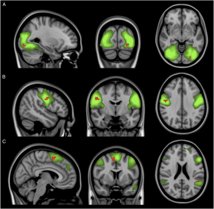
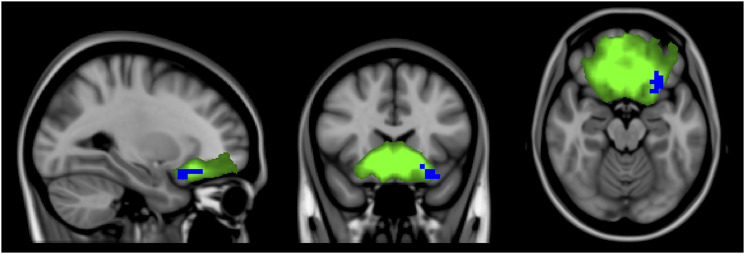
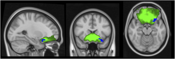
Results: In comparison to controls, the MS patients treated with natalizumab presented decreased connectivity in the left orbitofrontal cortex, in the anterior cingulate and orbitofrontal cortex network. The patients not treated with natalizumab presented increased connectivity in the secondary visual, sensorimotor, and ventral attention networks in comparison to controls.Compared to patients treated with natalizumab, the patients not using natalizumab presented increased connectivity in the left Heschl's gyrus and in the right superior frontal gyrus in the ventral attention network.
Conclusion: Differences in brain connectivity between MS patients not treated with natalizumab, healthy controls, and patients treated with natalizumab may be secondary to suboptimal neuronal compensation due to prior less efficient treatments, or due to a compensation in response to maladaptive plasticity.

 求助内容:
求助内容: 应助结果提醒方式:


