Katibe Tugce Temur, Guldane Magat, Melis Yılmaz, Sevgi Ozcan
{"title":"Evaluation of the effect of sickle cell disease on the mandibular bone of children and adolescents by image texture and radiomorphometric analysis.","authors":"Katibe Tugce Temur, Guldane Magat, Melis Yılmaz, Sevgi Ozcan","doi":"10.1007/s11282-023-00704-8","DOIUrl":null,"url":null,"abstract":"<p><strong>Objectives: </strong>Sickle cell disease (SCD) can cause osteoporotic changes in the jaw bones. In this study, it was aimed to evaluate possible bone changes using fractal analysis (FA) and morphometric analyses in dental panoramic radiographs of children and adolescents diagnosed with both homozygous and heterozygous forms of SCD.</p><p><strong>Methods: </strong>Sixty-five individuals (33 SCD, 32 controls) aged 6-17 years were included in the study. Four separate areas of interest (ROI) were selected for the right and left sides of all panoramic radiographs, and the FA value of the ROIs was calculated. Mandibular cortical width (MCW), panoramic mandibular index (PMI) and mandibular cortical index (MCI) and were evaluated. Data were statistically analyzed and p < 0.05 was accepted for statistical significance.</p><p><strong>Results: </strong>Fractal values of right and left ROI1 (the center of the mandibular angle.) and ROI4 (the cortical bone), and right ROI2 (the middle of the mandibular ramus) were statistically lower in the case group (p < 0.05). Right ROI2 and ROI4 fractal values of individuals in the case group were lower than those on the left side (p < 0.05). While MCI categories did not differ from the case-control group (p > 0.05), PMI and MCW values were lower in the case group (p < 0.05). All evaluated parameters did not differ according to age and gender (p > 0.05).</p><p><strong>Conclusion: </strong>The results of this study showed that SCD affects the mandible. FA, MCW and PMI parameters can be used to detect early osteoporotic changes in the disease.</p>","PeriodicalId":56103,"journal":{"name":"Oral Radiology","volume":null,"pages":null},"PeriodicalIF":1.6000,"publicationDate":"2023-10-01","publicationTypes":"Journal Article","fieldsOfStudy":null,"isOpenAccess":false,"openAccessPdf":"","citationCount":"0","resultStr":null,"platform":"Semanticscholar","paperid":null,"PeriodicalName":"Oral Radiology","FirstCategoryId":"3","ListUrlMain":"https://doi.org/10.1007/s11282-023-00704-8","RegionNum":3,"RegionCategory":"医学","ArticlePicture":[],"TitleCN":null,"AbstractTextCN":null,"PMCID":null,"EPubDate":"2023/8/3 0:00:00","PubModel":"Epub","JCR":"Q3","JCRName":"DENTISTRY, ORAL SURGERY & MEDICINE","Score":null,"Total":0}
引用次数: 0
Abstract
Objectives: Sickle cell disease (SCD) can cause osteoporotic changes in the jaw bones. In this study, it was aimed to evaluate possible bone changes using fractal analysis (FA) and morphometric analyses in dental panoramic radiographs of children and adolescents diagnosed with both homozygous and heterozygous forms of SCD.
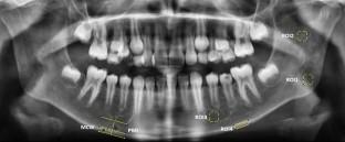
Methods: Sixty-five individuals (33 SCD, 32 controls) aged 6-17 years were included in the study. Four separate areas of interest (ROI) were selected for the right and left sides of all panoramic radiographs, and the FA value of the ROIs was calculated. Mandibular cortical width (MCW), panoramic mandibular index (PMI) and mandibular cortical index (MCI) and were evaluated. Data were statistically analyzed and p < 0.05 was accepted for statistical significance.
Results: Fractal values of right and left ROI1 (the center of the mandibular angle.) and ROI4 (the cortical bone), and right ROI2 (the middle of the mandibular ramus) were statistically lower in the case group (p < 0.05). Right ROI2 and ROI4 fractal values of individuals in the case group were lower than those on the left side (p < 0.05). While MCI categories did not differ from the case-control group (p > 0.05), PMI and MCW values were lower in the case group (p < 0.05). All evaluated parameters did not differ according to age and gender (p > 0.05).
Conclusion: The results of this study showed that SCD affects the mandible. FA, MCW and PMI parameters can be used to detect early osteoporotic changes in the disease.
期刊介绍:
As the official English-language journal of the Japanese Society for Oral and Maxillofacial Radiology and the Asian Academy of Oral and Maxillofacial Radiology, Oral Radiology is intended to be a forum for international collaboration in head and neck diagnostic imaging and all related fields. Oral Radiology features cutting-edge research papers, review articles, case reports, and technical notes from both the clinical and experimental fields. As membership in the Society is not a prerequisite, contributions are welcome from researchers and clinicians worldwide.

 求助内容:
求助内容: 应助结果提醒方式:


