{"title":"Corneal Ectasia after Laser-Assisted Small-Incision Lenticule Extraction: The Case for an Enhanced Ectasia Risk Assessment.","authors":"Siamak Zarei-Ghanavati, Samira Hassanzadeh, Renato Ambrósio","doi":"10.4103/joco.joco_79_22","DOIUrl":null,"url":null,"abstract":"<p><strong>Purpose: </strong>To present a case of asymmetric progressive corneal ectasia following femtosecond laser-assisted small-incision lenticule extraction.</p><p><strong>Methods: </strong>After obtaining a patient's consent, preoperative and postoperative findings were represented in this case report.</p><p><strong>Results: </strong>A 29-year-old woman presented with normal preoperative Placido disk-based corneal topography and tomographic findings. The corrected refractive error was -4.00 and -4.50 -1.00 × 177 in the right and left eye, respectively, with a maximal lenticule thickness of 87 and 115 μm OD/OS. Twenty months postoperatively, the patient presented with decreased vision in the left eye and mild ectatic changes in corneal shape in both eyes. The retrospective evaluation of the integrated rotating Scheimpflug tomography (Pentacam; Oculus, Wetzlar, Germany) and corneal biomechanical (Corvis ST) assessment revealed moderate susceptibility for corneal ectasia in the right eye and a significant corneal ectasia in the left eye.</p><p><strong>Conclusion: </strong>This case corroborates the need for an enhanced multimodal approach to characterize the risk for postoperative corneal ectasia after laser vision correction.</p>","PeriodicalId":15423,"journal":{"name":"Journal of Current Ophthalmology","volume":"34 3","pages":"357-363"},"PeriodicalIF":1.2000,"publicationDate":"2022-11-30","publicationTypes":"Journal Article","fieldsOfStudy":null,"isOpenAccess":false,"openAccessPdf":"https://ftp.ncbi.nlm.nih.gov/pub/pmc/oa_pdf/2d/b8/JCO-34-357.PMC9832456.pdf","citationCount":"0","resultStr":null,"platform":"Semanticscholar","paperid":null,"PeriodicalName":"Journal of Current Ophthalmology","FirstCategoryId":"1085","ListUrlMain":"https://doi.org/10.4103/joco.joco_79_22","RegionNum":0,"RegionCategory":null,"ArticlePicture":[],"TitleCN":null,"AbstractTextCN":null,"PMCID":null,"EPubDate":"2022/7/1 0:00:00","PubModel":"eCollection","JCR":"Q3","JCRName":"OPHTHALMOLOGY","Score":null,"Total":0}
引用次数: 0
Abstract
Purpose: To present a case of asymmetric progressive corneal ectasia following femtosecond laser-assisted small-incision lenticule extraction.
Methods: After obtaining a patient's consent, preoperative and postoperative findings were represented in this case report.
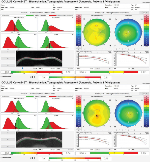
Results: A 29-year-old woman presented with normal preoperative Placido disk-based corneal topography and tomographic findings. The corrected refractive error was -4.00 and -4.50 -1.00 × 177 in the right and left eye, respectively, with a maximal lenticule thickness of 87 and 115 μm OD/OS. Twenty months postoperatively, the patient presented with decreased vision in the left eye and mild ectatic changes in corneal shape in both eyes. The retrospective evaluation of the integrated rotating Scheimpflug tomography (Pentacam; Oculus, Wetzlar, Germany) and corneal biomechanical (Corvis ST) assessment revealed moderate susceptibility for corneal ectasia in the right eye and a significant corneal ectasia in the left eye.
Conclusion: This case corroborates the need for an enhanced multimodal approach to characterize the risk for postoperative corneal ectasia after laser vision correction.
期刊介绍:
Peer Review under the responsibility of Iranian Society of Ophthalmology Journal of Current Ophthalmology, the official publication of the Iranian Society of Ophthalmology, is a peer-reviewed, open-access, scientific journal that welcomes high quality original articles related to vision science and all fields of ophthalmology. Journal of Current Ophthalmology is the continuum of Iranian Journal of Ophthalmology published since 1969.


 求助内容:
求助内容: 应助结果提醒方式:


