{"title":"Mechanism of sac expansion without evident endoleak analyzed with X ray phase-contrast tomography","authors":"Takateru Yamamoto MD , Takuro Tsukube MD, PhD , Yuko Wada MD, PhD , Masato Hoshino PhD , Naoto Yagi PhD , Kazunori Nakagawa PhD , Yutaka Nakashima MD, PhD , Kenji Okada MD, PhD , Tatsuichiro Seto MD, PhD","doi":"10.1016/j.jvssci.2023.100123","DOIUrl":null,"url":null,"abstract":"<div><h3>Objective</h3><p>Synchrotron radiation-based X ray phase-contrast tomography (XPCT) was used in this study to evaluate abdominal aorta specimens from patients with sac expansion without evidence of an endoleak (endotension) following endovascular aortic repair (EVAR) for an abdominal aortic aneurysm (AAA). The aim of this study was to analyze the morphologic structure of the aortic wall in patients with this condition and to establish the cause of the endotension.</p></div><div><h3>Methods</h3><p>Human aortic specimens of the abdominal aorta were obtained during open repair, fixed with formalin, and analyzed among three groups. Group A was specimens from open abdominal aortic aneurysm repairs (n = 7). Group E was specimens from sac expansion without an evident endoleak after EVAR (n = 7). Group N was specimens from non-aneurysmal “normal” cadaveric abdominal aortas (n = 5). Using XPCT (effective voxel size, 12.5 μm; density resolution, 1 mg/cm<sup>3</sup>), we measured the density of the tunica media (TM) in six regions of each sample. Then, any changes to the elastic lamina and the vasa vasorum were analyzed pathologically. The specimens were immunohistochemically examined with anti-CD31 and vascular endothelial growth factor antibodies.</p></div><div><h3>Results</h3><p>The time from EVAR to open aortic repair was 64.2 ± 7.2 months. There were significant differences in the thickness of the TM among three groups: 0.98 ± 0.03 mm in Group N; 0.31 ± 0.01 mm in Group A; and 0.15 ± 0.03 mm in Group E (<em>P</em> < .005). There were significant differences in the TM density among the groups: 1.087 ± 0.004 g/cm<sup>3</sup> in Group N; 1.070 ± 0.001 g/cm<sup>3</sup> in Group A; and 1.062 ± 0.007 g/cm<sup>3</sup> in Group E (<em>P</em> < .005). Differences in the thickness and density of the TM correlated with the thickness of the elastic lamina; in Group N, uniform high-density elastic fibers were observed in the TM. By contrast, a thinning of the elastic lamina in the TM was observed in Group A. A marked thinness and loss of elastic fibers was observed in Group E. CD31 immunostaining revealed that the vasa vasorum was localized in the adventitia and inside the outer third of the TM in Group N, and in the middle of the TM in Group A. In Group E, the vasa vasorum advanced up to the intima with vascular endothelial growth factor-positive cells in the intimal section.</p></div><div><h3>Conclusions</h3><p>XPCT could be used to demonstrate the densitometric property of the aortic aneurysmal wall after EVAR. We confirmed that the deformation process that occurs in the sac expansion after EVAR without evidence of an endoleak could be explained by hypoxia in the aortic wall.</p></div>","PeriodicalId":74035,"journal":{"name":"JVS-vascular science","volume":null,"pages":null},"PeriodicalIF":0.0000,"publicationDate":"2023-01-01","publicationTypes":"Journal Article","fieldsOfStudy":null,"isOpenAccess":false,"openAccessPdf":"https://ftp.ncbi.nlm.nih.gov/pub/pmc/oa_pdf/55/1f/main.PMC10474490.pdf","citationCount":"0","resultStr":null,"platform":"Semanticscholar","paperid":null,"PeriodicalName":"JVS-vascular science","FirstCategoryId":"1085","ListUrlMain":"https://www.sciencedirect.com/science/article/pii/S2666350323000275","RegionNum":0,"RegionCategory":null,"ArticlePicture":[],"TitleCN":null,"AbstractTextCN":null,"PMCID":null,"EPubDate":"","PubModel":"","JCR":"Q3","JCRName":"Medicine","Score":null,"Total":0}
引用次数: 0
Abstract
Objective
Synchrotron radiation-based X ray phase-contrast tomography (XPCT) was used in this study to evaluate abdominal aorta specimens from patients with sac expansion without evidence of an endoleak (endotension) following endovascular aortic repair (EVAR) for an abdominal aortic aneurysm (AAA). The aim of this study was to analyze the morphologic structure of the aortic wall in patients with this condition and to establish the cause of the endotension.
Methods
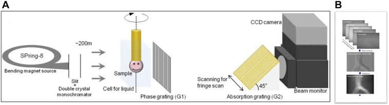
Human aortic specimens of the abdominal aorta were obtained during open repair, fixed with formalin, and analyzed among three groups. Group A was specimens from open abdominal aortic aneurysm repairs (n = 7). Group E was specimens from sac expansion without an evident endoleak after EVAR (n = 7). Group N was specimens from non-aneurysmal “normal” cadaveric abdominal aortas (n = 5). Using XPCT (effective voxel size, 12.5 μm; density resolution, 1 mg/cm3), we measured the density of the tunica media (TM) in six regions of each sample. Then, any changes to the elastic lamina and the vasa vasorum were analyzed pathologically. The specimens were immunohistochemically examined with anti-CD31 and vascular endothelial growth factor antibodies.
Results
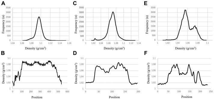
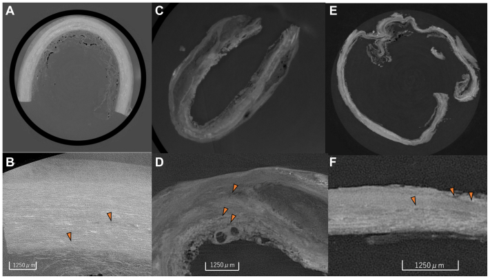
The time from EVAR to open aortic repair was 64.2 ± 7.2 months. There were significant differences in the thickness of the TM among three groups: 0.98 ± 0.03 mm in Group N; 0.31 ± 0.01 mm in Group A; and 0.15 ± 0.03 mm in Group E (P < .005). There were significant differences in the TM density among the groups: 1.087 ± 0.004 g/cm3 in Group N; 1.070 ± 0.001 g/cm3 in Group A; and 1.062 ± 0.007 g/cm3 in Group E (P < .005). Differences in the thickness and density of the TM correlated with the thickness of the elastic lamina; in Group N, uniform high-density elastic fibers were observed in the TM. By contrast, a thinning of the elastic lamina in the TM was observed in Group A. A marked thinness and loss of elastic fibers was observed in Group E. CD31 immunostaining revealed that the vasa vasorum was localized in the adventitia and inside the outer third of the TM in Group N, and in the middle of the TM in Group A. In Group E, the vasa vasorum advanced up to the intima with vascular endothelial growth factor-positive cells in the intimal section.
Conclusions
XPCT could be used to demonstrate the densitometric property of the aortic aneurysmal wall after EVAR. We confirmed that the deformation process that occurs in the sac expansion after EVAR without evidence of an endoleak could be explained by hypoxia in the aortic wall.

 求助内容:
求助内容: 应助结果提醒方式:


