Differences in the Effects of Pentobarbital Anesthetic and Combination of Medetomidine Hydrochloride, Midazolam, and Butorphanol Tartrate Anesthetic on Electroretinogram in Spontaneously Diabetic Torii Fatty Rats.
{"title":"Differences in the Effects of Pentobarbital Anesthetic and Combination of Medetomidine Hydrochloride, Midazolam, and Butorphanol Tartrate Anesthetic on Electroretinogram in Spontaneously Diabetic Torii Fatty Rats.","authors":"Tetsuya Hasegawa, Rina Takagi, Yoshiaki Tanaka, Takeshi Ohta, Masami Shinohara, Yasushi Kageyama, Tomohiko Sasase, Shin-Ichi Muramatsu, Toshikatsu Kaburaki, Akihiro Kakehashi","doi":"10.1159/000526189","DOIUrl":null,"url":null,"abstract":"<p><strong>Purpose: </strong>The aim of this study was to investigate the effects of different anesthetic agents on electroretinograms (ERGs) in Spontaneously Diabetic Torii fatty rats (SDT fatty rats).</p><p><strong>Methods: </strong>The ERG recordings were measured under general anesthesia using pentobarbital or a combination of medetomidine hydrochloride, midazolam, and butorphanol (MMB) tartrate anesthesia in 12 9-week-old normal Sprague-Dawley rats (Jcl:SD rats) and 16 SDT fatty rats. Each animal model was divided into 2 groups, the pentobarbital group and MMB group. The amplitudes and peak times of the a- and b-waves and oscillatory potentials (OPs) were measured from 0.0001 candela per square meter (cd.s/m<sup>2</sup>) to 10.0 cd.s/m<sup>2</sup>.</p><p><strong>Results: </strong>The amplitude of the a-wave was significantly higher in the MMB group of Jcl:SD rats, but there was no significant difference in amplitude between the two groups of SDT fatty rats. There was no significant difference in the OP1 amplitude between both groups of Jcl:SD rats, but the OP1 amplitude was significantly higher in the MMB group of SDT fatty rats. The OP2 amplitude was significantly higher in the pentobarbital group in both the Jcl:SD rats and SDT fatty rats. There was no significant difference in the OP3 amplitude between the Jcl:SD and SDT fatty rat groups. The amplitude of the OP4 waves was significantly higher in the MMB group for both Jcl:SD and SDT fatty rats. There was no significant difference in the sums of the OP1 to OP4 (ΣOPs) amplitudes between the Jcl:SD and SDT fatty rat groups. There was no significant difference in the b-wave amplitude between the Jcl:SD rat groups, but the b-wave amplitude was significantly higher in the SDT fatty rats that received pentobarbital. The peak times for a-wave, OP1, OP2, OP3, OP4, and ΣOPs were significantly longer in the pentobarbital group of SD rats. The peak time of the b-wave was significantly longer in the MMB group of Jcl:SD rats, but the same result was obtained in the SDT fatty rats except that there was no significant difference in the a-wave.</p><p><strong>Conclusion: </strong>The overall ERG results vary depending on the anesthetic agent used. The OPs can be observed in detail when using MMB. Since the SDT fatty rat is a diabetic model animal, we recommend MMB as the anesthesia of choice when studying the OP waves in detail.</p>","PeriodicalId":9075,"journal":{"name":"Biomedicine Hub","volume":"7 3","pages":"106-114"},"PeriodicalIF":0.0000,"publicationDate":"2022-09-01","publicationTypes":"Journal Article","fieldsOfStudy":null,"isOpenAccess":false,"openAccessPdf":"https://ftp.ncbi.nlm.nih.gov/pub/pmc/oa_pdf/40/2f/bmh-0007-0106.PMC9574210.pdf","citationCount":"1","resultStr":null,"platform":"Semanticscholar","paperid":null,"PeriodicalName":"Biomedicine Hub","FirstCategoryId":"1085","ListUrlMain":"https://doi.org/10.1159/000526189","RegionNum":0,"RegionCategory":null,"ArticlePicture":[],"TitleCN":null,"AbstractTextCN":null,"PMCID":null,"EPubDate":"","PubModel":"","JCR":"","JCRName":"","Score":null,"Total":0}
引用次数: 1
Abstract
Purpose: The aim of this study was to investigate the effects of different anesthetic agents on electroretinograms (ERGs) in Spontaneously Diabetic Torii fatty rats (SDT fatty rats).
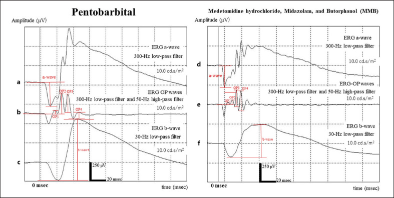
Methods: The ERG recordings were measured under general anesthesia using pentobarbital or a combination of medetomidine hydrochloride, midazolam, and butorphanol (MMB) tartrate anesthesia in 12 9-week-old normal Sprague-Dawley rats (Jcl:SD rats) and 16 SDT fatty rats. Each animal model was divided into 2 groups, the pentobarbital group and MMB group. The amplitudes and peak times of the a- and b-waves and oscillatory potentials (OPs) were measured from 0.0001 candela per square meter (cd.s/m2) to 10.0 cd.s/m2.
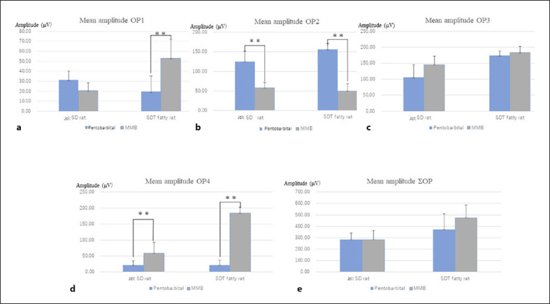
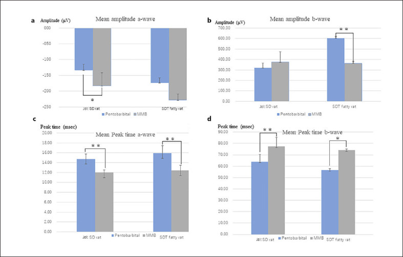
Results: The amplitude of the a-wave was significantly higher in the MMB group of Jcl:SD rats, but there was no significant difference in amplitude between the two groups of SDT fatty rats. There was no significant difference in the OP1 amplitude between both groups of Jcl:SD rats, but the OP1 amplitude was significantly higher in the MMB group of SDT fatty rats. The OP2 amplitude was significantly higher in the pentobarbital group in both the Jcl:SD rats and SDT fatty rats. There was no significant difference in the OP3 amplitude between the Jcl:SD and SDT fatty rat groups. The amplitude of the OP4 waves was significantly higher in the MMB group for both Jcl:SD and SDT fatty rats. There was no significant difference in the sums of the OP1 to OP4 (ΣOPs) amplitudes between the Jcl:SD and SDT fatty rat groups. There was no significant difference in the b-wave amplitude between the Jcl:SD rat groups, but the b-wave amplitude was significantly higher in the SDT fatty rats that received pentobarbital. The peak times for a-wave, OP1, OP2, OP3, OP4, and ΣOPs were significantly longer in the pentobarbital group of SD rats. The peak time of the b-wave was significantly longer in the MMB group of Jcl:SD rats, but the same result was obtained in the SDT fatty rats except that there was no significant difference in the a-wave.
Conclusion: The overall ERG results vary depending on the anesthetic agent used. The OPs can be observed in detail when using MMB. Since the SDT fatty rat is a diabetic model animal, we recommend MMB as the anesthesia of choice when studying the OP waves in detail.

 求助内容:
求助内容: 应助结果提醒方式:


