{"title":"Reconstitution of Fusion-Competent Human Placental Fusogen Syncytin-2.","authors":"Lu Xu, Sha Sun","doi":"10.1007/s00232-022-00242-0","DOIUrl":null,"url":null,"abstract":"<p><p>Mammalian placenta formation requires continuous fusion of trophoblasts. Human endogenous retrovirus-derived proteins syncytin-1 and syncytin-2 mediate cell-cell fusion of placental cytotrophoblasts to form syncytiotrophoblasts in primates, which is required for normal placenta function and fetal development. Syncytins are post-translationally cleaved by the endoprotease furin into surface (SU) and transmembrane (TM) subunits for activation. Little is currently known about the molecular mechanisms of syncytin-mediated cell-cell fusion, and their functions have not been well studied in vitro. Here, we express tagged syncytin-2 in mammalian HEK293T cells and demonstrate that the tagging greatly influences the cleavage and fusogenic activity of syncytin-2. By detecting the N-terminal tagged SU, we find that it is released into the extracellular space during the fusion process. Furthermore, when N-linked glycosylation and disulfide bond formation are blocked, the cleavage and fusogenic activity of syncytin-2 are inhibited. Finally, we were able to purify functional syncytin-2 from HEK293T cells and incorporate it into proteoliposomes. These findings lay a solid foundation for interogating the molecular mechanisms of syncytin-2-mediated cell-cell fusion in vitro.</p>","PeriodicalId":2,"journal":{"name":"ACS Applied Bio Materials","volume":null,"pages":null},"PeriodicalIF":4.6000,"publicationDate":"2022-12-01","publicationTypes":"Journal Article","fieldsOfStudy":null,"isOpenAccess":false,"openAccessPdf":"","citationCount":"1","resultStr":null,"platform":"Semanticscholar","paperid":null,"PeriodicalName":"ACS Applied Bio Materials","FirstCategoryId":"99","ListUrlMain":"https://doi.org/10.1007/s00232-022-00242-0","RegionNum":0,"RegionCategory":null,"ArticlePicture":[],"TitleCN":null,"AbstractTextCN":null,"PMCID":null,"EPubDate":"","PubModel":"","JCR":"Q2","JCRName":"MATERIALS SCIENCE, BIOMATERIALS","Score":null,"Total":0}
引用次数: 1
Abstract
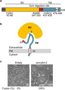
Mammalian placenta formation requires continuous fusion of trophoblasts. Human endogenous retrovirus-derived proteins syncytin-1 and syncytin-2 mediate cell-cell fusion of placental cytotrophoblasts to form syncytiotrophoblasts in primates, which is required for normal placenta function and fetal development. Syncytins are post-translationally cleaved by the endoprotease furin into surface (SU) and transmembrane (TM) subunits for activation. Little is currently known about the molecular mechanisms of syncytin-mediated cell-cell fusion, and their functions have not been well studied in vitro. Here, we express tagged syncytin-2 in mammalian HEK293T cells and demonstrate that the tagging greatly influences the cleavage and fusogenic activity of syncytin-2. By detecting the N-terminal tagged SU, we find that it is released into the extracellular space during the fusion process. Furthermore, when N-linked glycosylation and disulfide bond formation are blocked, the cleavage and fusogenic activity of syncytin-2 are inhibited. Finally, we were able to purify functional syncytin-2 from HEK293T cells and incorporate it into proteoliposomes. These findings lay a solid foundation for interogating the molecular mechanisms of syncytin-2-mediated cell-cell fusion in vitro.
 求助内容:
求助内容: 应助结果提醒方式:


