Jelena Levi, Heying Duan, Shahriar Yaghoubi, Juliet Packiasamy, Lyna Huynh, Tina Lam, Faiq Shaikh, Deepak Behera, Hong Song, Joseph Blecha, Salma Jivan, Youngho Seo, Henry F VanBrocklin
{"title":"Biodistribution of a Mitochondrial Metabolic Tracer, [<sup>18</sup>F]F-AraG, in Healthy Volunteers.","authors":"Jelena Levi, Heying Duan, Shahriar Yaghoubi, Juliet Packiasamy, Lyna Huynh, Tina Lam, Faiq Shaikh, Deepak Behera, Hong Song, Joseph Blecha, Salma Jivan, Youngho Seo, Henry F VanBrocklin","doi":"10.1155/2022/3667417","DOIUrl":null,"url":null,"abstract":"<p><strong>Purpose: </strong>[<sup>18</sup>F]F-AraG is a radiolabeled nucleoside analog that shows relative specificity for activated T cells. The aim of this study was to investigate the biodistribution of [<sup>18</sup>F]F-AraG in healthy volunteers and assess the preliminary safety and radiation dosimetry.</p><p><strong>Methods: </strong>Six healthy subjects (three female and three male) between the ages of 24 and 60 participated in the study. Each subject received a bolus venous injection of [<sup>18</sup>F]F-AraG (dose range: 244.2-329.3 MBq) prior to four consecutive PET/MR whole-body scans. Blood samples were collected at regular intervals and vital signs monitored before and after tracer administration. Regions of interest were delineated for multiple organs, and the area under the time-activity curves was calculated for each organ and used to derive time-integrated activity coefficient (TIAC). TIACs were input for absorbed dose and effective dose calculations using OLINDA.</p><p><strong>Results: </strong>PET/MR examination was well tolerated, and no adverse effects to the administration of [<sup>18</sup>F]F-AraG were noted by the study participants. The biodistribution was generally reflective of the expression and activity profiles of the enzymes involved in [<sup>18</sup>F]F-AraG's cellular accumulation, mitochondrial kinase dGK, and SAMHD1. The highest uptake was observed in the kidneys and liver, while the brain, lung, bone marrow, and muscle showed low tracer uptake. The estimated effective dose for [<sup>18</sup>F]F-AraG was 0.0162 mSv/MBq (0.0167 mSv/MBq for females and 0.0157 mSv/MBq for males).</p><p><strong>Conclusion: </strong>Biodistribution of [<sup>18</sup>F]F-AraG in healthy volunteers was consistent with its association with mitochondrial metabolism. PET/MR [<sup>18</sup>F]F-AraG imaging was well tolerated, with a radiation dosimetry profile similar to other commonly used [<sup>18</sup>F]-labeled tracers. [<sup>18</sup>F]F-AraG's connection with mitochondrial biogenesis and favorable biodistribution characteristics make it an attractive tracer with a variety of potential applications.</p>","PeriodicalId":18855,"journal":{"name":"Molecular Imaging","volume":"2022 ","pages":"3667417"},"PeriodicalIF":2.4000,"publicationDate":"2022-01-01","publicationTypes":"Journal Article","fieldsOfStudy":null,"isOpenAccess":false,"openAccessPdf":"https://www.ncbi.nlm.nih.gov/pmc/articles/PMC9400547/pdf/","citationCount":"5","resultStr":null,"platform":"Semanticscholar","paperid":null,"PeriodicalName":"Molecular Imaging","FirstCategoryId":"3","ListUrlMain":"https://doi.org/10.1155/2022/3667417","RegionNum":4,"RegionCategory":"医学","ArticlePicture":[],"TitleCN":null,"AbstractTextCN":null,"PMCID":null,"EPubDate":"","PubModel":"","JCR":"Q3","JCRName":"BIOCHEMICAL RESEARCH METHODS","Score":null,"Total":0}
引用次数: 5
Abstract
Purpose: [18F]F-AraG is a radiolabeled nucleoside analog that shows relative specificity for activated T cells. The aim of this study was to investigate the biodistribution of [18F]F-AraG in healthy volunteers and assess the preliminary safety and radiation dosimetry.
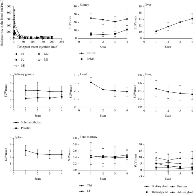
Methods: Six healthy subjects (three female and three male) between the ages of 24 and 60 participated in the study. Each subject received a bolus venous injection of [18F]F-AraG (dose range: 244.2-329.3 MBq) prior to four consecutive PET/MR whole-body scans. Blood samples were collected at regular intervals and vital signs monitored before and after tracer administration. Regions of interest were delineated for multiple organs, and the area under the time-activity curves was calculated for each organ and used to derive time-integrated activity coefficient (TIAC). TIACs were input for absorbed dose and effective dose calculations using OLINDA.
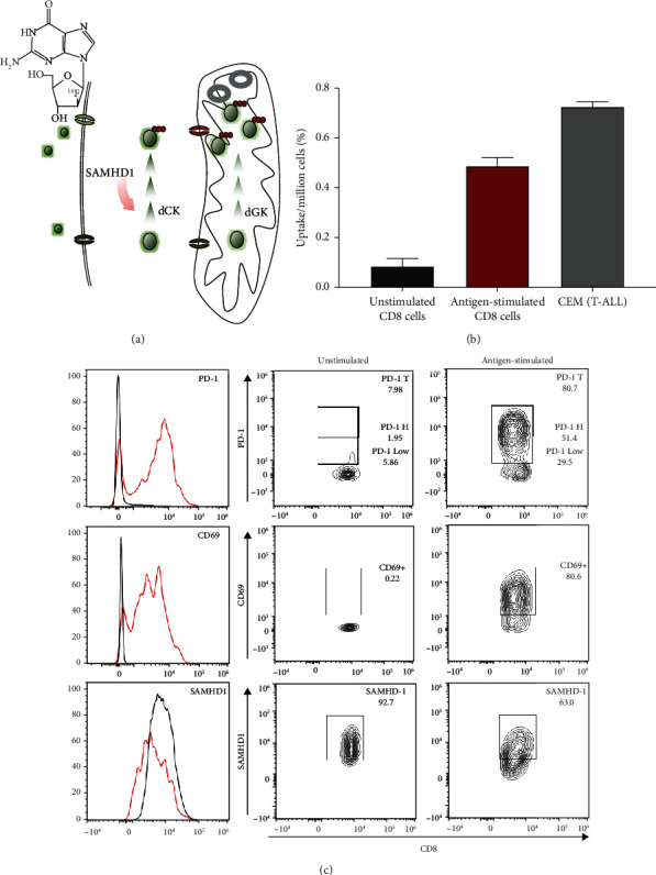
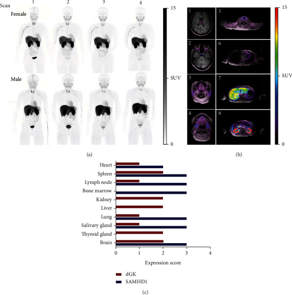
Results: PET/MR examination was well tolerated, and no adverse effects to the administration of [18F]F-AraG were noted by the study participants. The biodistribution was generally reflective of the expression and activity profiles of the enzymes involved in [18F]F-AraG's cellular accumulation, mitochondrial kinase dGK, and SAMHD1. The highest uptake was observed in the kidneys and liver, while the brain, lung, bone marrow, and muscle showed low tracer uptake. The estimated effective dose for [18F]F-AraG was 0.0162 mSv/MBq (0.0167 mSv/MBq for females and 0.0157 mSv/MBq for males).
Conclusion: Biodistribution of [18F]F-AraG in healthy volunteers was consistent with its association with mitochondrial metabolism. PET/MR [18F]F-AraG imaging was well tolerated, with a radiation dosimetry profile similar to other commonly used [18F]-labeled tracers. [18F]F-AraG's connection with mitochondrial biogenesis and favorable biodistribution characteristics make it an attractive tracer with a variety of potential applications.
Molecular ImagingBiochemistry, Genetics and Molecular Biology-Biotechnology
自引率
3.60%
发文量
21
期刊介绍:
Molecular Imaging is a peer-reviewed, open access journal highlighting the breadth of molecular imaging research from basic science to preclinical studies to human applications. This serves both the scientific and clinical communities by disseminating novel results and concepts relevant to the biological study of normal and disease processes in both basic and translational studies ranging from mice to humans.
 求助内容:
求助内容: 应助结果提醒方式:


