Faisal Shaikh, Jon-Emile Kenny, Omar Awan, Daniela Markovic, Oren Friedman, Tao He, Sidharth Singh, Peter Yan, Nida Qadir, Igor Barjaktarevic
{"title":"Measuring the accuracy of cardiac output using POCUS: the introduction of artificial intelligence into routine care.","authors":"Faisal Shaikh, Jon-Emile Kenny, Omar Awan, Daniela Markovic, Oren Friedman, Tao He, Sidharth Singh, Peter Yan, Nida Qadir, Igor Barjaktarevic","doi":"10.1186/s13089-022-00301-6","DOIUrl":null,"url":null,"abstract":"<p><strong>Background: </strong>Shock management requires quick and reliable means to monitor the hemodynamic effects of fluid resuscitation. Point-of-care ultrasound (POCUS) is a relatively quick and non-invasive imaging technique capable of capturing cardiac output (CO) variations in acute settings. However, POCUS is plagued by variable operator skill and interpretation. Artificial intelligence may assist healthcare professionals obtain more objective and precise measurements during ultrasound imaging, thus increasing usability among users with varying experience. In this feasibility study, we compared the performance of novice POCUS users in measuring CO with manual techniques to a novel automation-assisted technique that provides real-time feedback to correct image acquisition for optimal aortic outflow velocity measurement.</p><p><strong>Methods: </strong>28 junior critical care trainees with limited experience in POCUS performed manual and automation-assisted CO measurements on a single healthy volunteer. CO measurements were obtained using left ventricular outflow tract (LVOT) velocity time integral (VTI) and LVOT diameter. Measurements obtained by study subjects were compared to those taken by board-certified echocardiographers. Comparative analyses were performed using Spearman's rank correlation and Bland-Altman matched-pairs analysis.</p><p><strong>Results: </strong>Adequate image acquisition was 100% feasible. The correlation between manual and automated VTI values was not significant (p = 0.11) and means from both groups underestimated the mean values obtained by board-certified echocardiographers. Automated measurements of VTI in the trainee cohort were found to have more reproducibility, narrower measurement range (6.2 vs. 10.3 cm), and reduced standard deviation (1.98 vs. 2.33 cm) compared to manual measurements. The coefficient of variation across raters was 11.5%, 13.6% and 15.4% for board-certified echocardiographers, automated, and manual VTI tracing, respectively.</p><p><strong>Conclusions: </strong>Our study demonstrates that novel automation-assisted VTI is feasible and can decrease variability while increasing precision in CO measurement. These results support the use of artificial intelligence-augmented image acquisition in routine critical care ultrasound and may have a role for evaluating the response of CO to hemodynamic interventions. Further investigations into artificial intelligence-assisted ultrasound systems in clinical settings are warranted.</p>","PeriodicalId":36911,"journal":{"name":"Ultrasound Journal","volume":null,"pages":null},"PeriodicalIF":3.4000,"publicationDate":"2022-12-14","publicationTypes":"Journal Article","fieldsOfStudy":null,"isOpenAccess":false,"openAccessPdf":"https://www.ncbi.nlm.nih.gov/pmc/articles/PMC9751239/pdf/","citationCount":"3","resultStr":null,"platform":"Semanticscholar","paperid":null,"PeriodicalName":"Ultrasound Journal","FirstCategoryId":"1085","ListUrlMain":"https://doi.org/10.1186/s13089-022-00301-6","RegionNum":0,"RegionCategory":null,"ArticlePicture":[],"TitleCN":null,"AbstractTextCN":null,"PMCID":null,"EPubDate":"","PubModel":"","JCR":"Q2","JCRName":"Medicine","Score":null,"Total":0}
引用次数: 3
Abstract
Background: Shock management requires quick and reliable means to monitor the hemodynamic effects of fluid resuscitation. Point-of-care ultrasound (POCUS) is a relatively quick and non-invasive imaging technique capable of capturing cardiac output (CO) variations in acute settings. However, POCUS is plagued by variable operator skill and interpretation. Artificial intelligence may assist healthcare professionals obtain more objective and precise measurements during ultrasound imaging, thus increasing usability among users with varying experience. In this feasibility study, we compared the performance of novice POCUS users in measuring CO with manual techniques to a novel automation-assisted technique that provides real-time feedback to correct image acquisition for optimal aortic outflow velocity measurement.
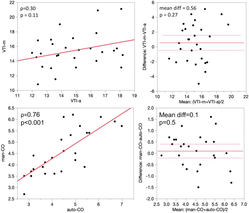
Methods: 28 junior critical care trainees with limited experience in POCUS performed manual and automation-assisted CO measurements on a single healthy volunteer. CO measurements were obtained using left ventricular outflow tract (LVOT) velocity time integral (VTI) and LVOT diameter. Measurements obtained by study subjects were compared to those taken by board-certified echocardiographers. Comparative analyses were performed using Spearman's rank correlation and Bland-Altman matched-pairs analysis.
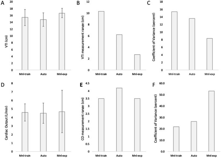
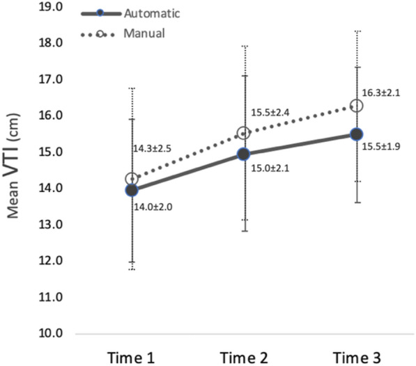
Results: Adequate image acquisition was 100% feasible. The correlation between manual and automated VTI values was not significant (p = 0.11) and means from both groups underestimated the mean values obtained by board-certified echocardiographers. Automated measurements of VTI in the trainee cohort were found to have more reproducibility, narrower measurement range (6.2 vs. 10.3 cm), and reduced standard deviation (1.98 vs. 2.33 cm) compared to manual measurements. The coefficient of variation across raters was 11.5%, 13.6% and 15.4% for board-certified echocardiographers, automated, and manual VTI tracing, respectively.
Conclusions: Our study demonstrates that novel automation-assisted VTI is feasible and can decrease variability while increasing precision in CO measurement. These results support the use of artificial intelligence-augmented image acquisition in routine critical care ultrasound and may have a role for evaluating the response of CO to hemodynamic interventions. Further investigations into artificial intelligence-assisted ultrasound systems in clinical settings are warranted.

 求助内容:
求助内容: 应助结果提醒方式:


