Li-Jun Wu, Wei Lin, Jian-Jiang Liu, Wei-Xin Chen, Wen-Jun He, Yuan Shi, Xiao Liu, Ke Li
{"title":"Transplantation of human induced pluripotent stem cell derived keratinocytes accelerates deep second-degree burn wound healing.","authors":"Li-Jun Wu, Wei Lin, Jian-Jiang Liu, Wei-Xin Chen, Wen-Jun He, Yuan Shi, Xiao Liu, Ke Li","doi":"10.4252/wjsc.v15.i7.713","DOIUrl":null,"url":null,"abstract":"<p><strong>Background: </strong>Current evidence shows that human induced pluripotent stem cells (hiPSCs) can effectively differentiate into keratinocytes (KCs), but its effect on skin burn healing has not been reported.</p><p><strong>Aim: </strong>To observe the effects of hiPSCs-derived KCs transplantation on skin burn healing in mice and to preliminarily reveal the underlying mechanisms.</p><p><strong>Methods: </strong>An analysis of differentially expressed genes in burn wounds based on GEO datasets GSE140926, and GSE27186 was established. A differentiation medium containing retinoic acid and bone morphogenetic protein 4 was applied to induce hiPSCs to differentiate into KCs. The expression of KCs marker proteins was detected using immunofluorescence staining. A model of a C57BL/6 mouse with deep cutaneous second-degree burn was created, and then phosphate buffered saline (PBS), hiPSCs-KCs, or hiPSCs-KCs with knockdown of <i>COL7A1</i> were injected around the wound surface. The wound healing, re-epithelialization, engraftment of hiPSCs-KCs into wounds, proinflammatory factor level, and the NF-κB pathway proteins were assessed by hematoxylin-eosin staining, carboxifluorescein diacetate succinimidyl ester (CFSE) fluorescence staining, enzyme linked immunosorbent assay, and Western blotting on days 3, 7, and 14 after the injection, respectively. Moreover, the effects of <i>COL7A1</i> knockdown on the proliferation and migration of hiPSCs-KCs were confirmed by immunohistochemistry, EdU, Transwell, and damage repair assays.</p><p><strong>Results: </strong>HiPSCs-KCs could express the hallmark proteins of KCs. <i>COL7A1</i> was down-regulated in burn wound tissues and highly expressed in hiPSCs-KCs. Transplantation of hiPSCs-KCs into mice with burn wounds resulted in a significant decrease in wound area, an increase in wound re-epithelialization, a decrease in proinflammatory factors content, and an inhibition of NF-κB pathway activation compared to the PBS group. The in vitro assay showed that <i>COL7A1</i> knockdown could rescue the inhibition of hiPSCs-KCs proliferation and migration, providing further evidence that <i>COL7A1</i> speeds up burn wound healing by limiting cell proliferation and migration.</p><p><strong>Conclusion: </strong>In deep, second-degree burn wounds, <i>COL7A1</i> can promote KC proliferation and migration while also suppressing the inflammatory response.</p>","PeriodicalId":23775,"journal":{"name":"World journal of stem cells","volume":null,"pages":null},"PeriodicalIF":3.6000,"publicationDate":"2023-07-26","publicationTypes":"Journal Article","fieldsOfStudy":null,"isOpenAccess":false,"openAccessPdf":"https://ftp.ncbi.nlm.nih.gov/pub/pmc/oa_pdf/2e/c8/WJSC-15-713.PMC10401420.pdf","citationCount":"3","resultStr":null,"platform":"Semanticscholar","paperid":null,"PeriodicalName":"World journal of stem cells","FirstCategoryId":"3","ListUrlMain":"https://doi.org/10.4252/wjsc.v15.i7.713","RegionNum":3,"RegionCategory":"医学","ArticlePicture":[],"TitleCN":null,"AbstractTextCN":null,"PMCID":null,"EPubDate":"","PubModel":"","JCR":"Q3","JCRName":"CELL & TISSUE ENGINEERING","Score":null,"Total":0}
引用次数: 3
Abstract
Background: Current evidence shows that human induced pluripotent stem cells (hiPSCs) can effectively differentiate into keratinocytes (KCs), but its effect on skin burn healing has not been reported.
Aim: To observe the effects of hiPSCs-derived KCs transplantation on skin burn healing in mice and to preliminarily reveal the underlying mechanisms.
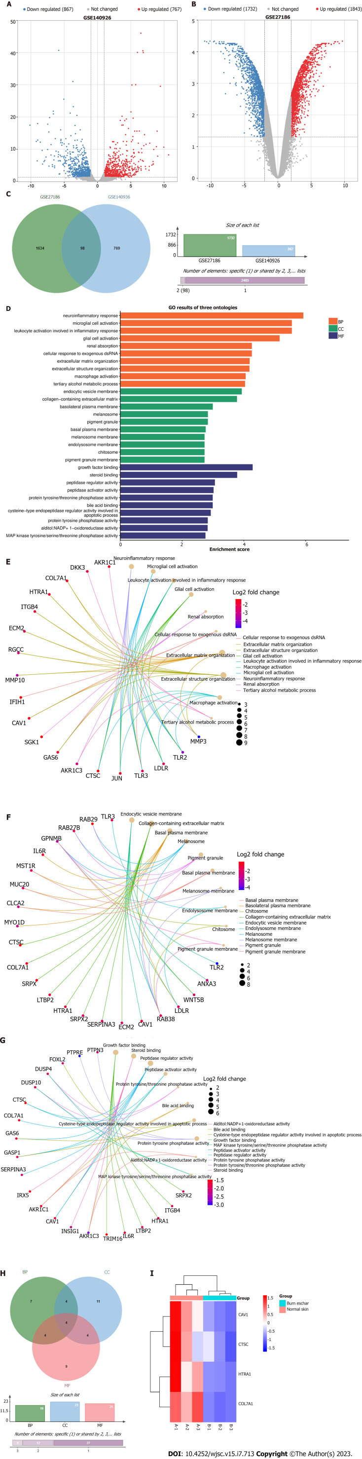
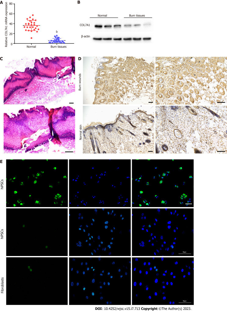
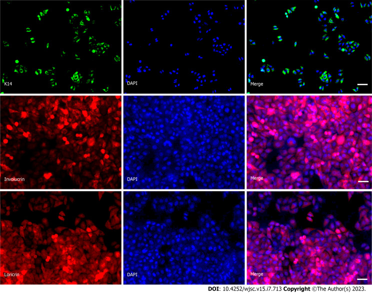
Methods: An analysis of differentially expressed genes in burn wounds based on GEO datasets GSE140926, and GSE27186 was established. A differentiation medium containing retinoic acid and bone morphogenetic protein 4 was applied to induce hiPSCs to differentiate into KCs. The expression of KCs marker proteins was detected using immunofluorescence staining. A model of a C57BL/6 mouse with deep cutaneous second-degree burn was created, and then phosphate buffered saline (PBS), hiPSCs-KCs, or hiPSCs-KCs with knockdown of COL7A1 were injected around the wound surface. The wound healing, re-epithelialization, engraftment of hiPSCs-KCs into wounds, proinflammatory factor level, and the NF-κB pathway proteins were assessed by hematoxylin-eosin staining, carboxifluorescein diacetate succinimidyl ester (CFSE) fluorescence staining, enzyme linked immunosorbent assay, and Western blotting on days 3, 7, and 14 after the injection, respectively. Moreover, the effects of COL7A1 knockdown on the proliferation and migration of hiPSCs-KCs were confirmed by immunohistochemistry, EdU, Transwell, and damage repair assays.
Results: HiPSCs-KCs could express the hallmark proteins of KCs. COL7A1 was down-regulated in burn wound tissues and highly expressed in hiPSCs-KCs. Transplantation of hiPSCs-KCs into mice with burn wounds resulted in a significant decrease in wound area, an increase in wound re-epithelialization, a decrease in proinflammatory factors content, and an inhibition of NF-κB pathway activation compared to the PBS group. The in vitro assay showed that COL7A1 knockdown could rescue the inhibition of hiPSCs-KCs proliferation and migration, providing further evidence that COL7A1 speeds up burn wound healing by limiting cell proliferation and migration.
Conclusion: In deep, second-degree burn wounds, COL7A1 can promote KC proliferation and migration while also suppressing the inflammatory response.
期刊介绍:
The World Journal of Stem Cells (WJSC) is a leading academic journal devoted to reporting the latest, cutting-edge research progress and findings of basic research and clinical practice in the field of stem cells. It was launched on December 31, 2009 and is published monthly (12 issues annually) by BPG, the world''s leading professional clinical medical journal publishing company.
 求助内容:
求助内容: 应助结果提醒方式:


