A bioscaffold of decellularized whole osteochondral sheet improves proliferation and differentiation of loaded mesenchymal stem cells in a rabbit model.
{"title":"A bioscaffold of decellularized whole osteochondral sheet improves proliferation and differentiation of loaded mesenchymal stem cells in a rabbit model.","authors":"Leila Taghiyar, Hamideh Asadi, Mohamadreza Baghaban Eslaminejad","doi":"10.1007/s10561-023-10084-2","DOIUrl":null,"url":null,"abstract":"<p><p>As a Natural decellularized extracellular matrix, osteochondral tissue is the best scaffold for the restoration of osteoarthritis defects. Bioscaffolds have the most similarly innate properties like biomechanical properties and the preserved connection of the bone-to-cartilage border. Although, their compacity and low porosity particularly, are proven to be difficulties of decellularization and cell penetration. This study aims to develop a new bioscaffold of decellularized osteochondral tissue (DOT) that is recellularized by bone marrow-derived mesenchymal stem cells (BM-MSCs), as a biphasic allograft, which preserved the interface between the cartilage section and subchondral bone of the joint. Whole osteochondral tissues of rabbit knee joints were sheeted in cartilaginous parts in 200-250 µm sections while connected to the subchondral bone and then fully decellularized. The BM-MSCs were seeded on the scaffolds in vitro; some constructs were subcutaneously implanted into the back of the rabbit. The cell penetration, differentiation to bone and cartilage, viability, and cell proliferation in vitro and in vivo were evaluated by qPCR, histological staining, MTT assay, and immunohistochemistry. DNA content analysis and SEM assessments confirmed the decellularization of the bioscaffold. Then, histological and SEM evaluations indicated that the cells could successfully penetrate the bone and cartilage lacunas in implanted grafts. MTT assay confirmed cell proliferation. Prominently, gene expression analysis showed that seeded cells differentiated into osteoblasts and chondrocytes in both bone and cartilage sections. More importantly, seeded cells on the bioscaffold started ECM secretion. Our results indicate that cartilage-to-bone border integrity was largely preserved. Additionally, ECM-sheeted DOT could be employed as a useful scaffold for promoting the regeneration of osteochondral defects.</p>","PeriodicalId":9723,"journal":{"name":"Cell and Tissue Banking","volume":" ","pages":"711-724"},"PeriodicalIF":1.4000,"publicationDate":"2023-12-01","publicationTypes":"Journal Article","fieldsOfStudy":null,"isOpenAccess":false,"openAccessPdf":"","citationCount":"1","resultStr":null,"platform":"Semanticscholar","paperid":null,"PeriodicalName":"Cell and Tissue Banking","FirstCategoryId":"5","ListUrlMain":"https://doi.org/10.1007/s10561-023-10084-2","RegionNum":4,"RegionCategory":"医学","ArticlePicture":[],"TitleCN":null,"AbstractTextCN":null,"PMCID":null,"EPubDate":"2023/3/20 0:00:00","PubModel":"Epub","JCR":"Q4","JCRName":"CELL BIOLOGY","Score":null,"Total":0}
引用次数: 1
Abstract
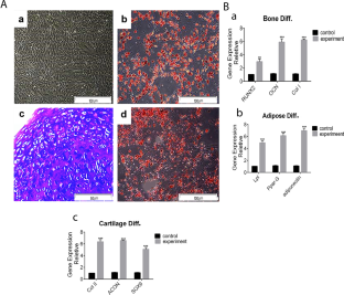
As a Natural decellularized extracellular matrix, osteochondral tissue is the best scaffold for the restoration of osteoarthritis defects. Bioscaffolds have the most similarly innate properties like biomechanical properties and the preserved connection of the bone-to-cartilage border. Although, their compacity and low porosity particularly, are proven to be difficulties of decellularization and cell penetration. This study aims to develop a new bioscaffold of decellularized osteochondral tissue (DOT) that is recellularized by bone marrow-derived mesenchymal stem cells (BM-MSCs), as a biphasic allograft, which preserved the interface between the cartilage section and subchondral bone of the joint. Whole osteochondral tissues of rabbit knee joints were sheeted in cartilaginous parts in 200-250 µm sections while connected to the subchondral bone and then fully decellularized. The BM-MSCs were seeded on the scaffolds in vitro; some constructs were subcutaneously implanted into the back of the rabbit. The cell penetration, differentiation to bone and cartilage, viability, and cell proliferation in vitro and in vivo were evaluated by qPCR, histological staining, MTT assay, and immunohistochemistry. DNA content analysis and SEM assessments confirmed the decellularization of the bioscaffold. Then, histological and SEM evaluations indicated that the cells could successfully penetrate the bone and cartilage lacunas in implanted grafts. MTT assay confirmed cell proliferation. Prominently, gene expression analysis showed that seeded cells differentiated into osteoblasts and chondrocytes in both bone and cartilage sections. More importantly, seeded cells on the bioscaffold started ECM secretion. Our results indicate that cartilage-to-bone border integrity was largely preserved. Additionally, ECM-sheeted DOT could be employed as a useful scaffold for promoting the regeneration of osteochondral defects.
期刊介绍:
Cell and Tissue Banking provides a forum for disseminating information to scientists and clinicians involved in the banking and transplantation of cells and tissues. Cell and Tissue Banking is an international, peer-reviewed journal that publishes original papers in the following areas:
basic research concerning general aspects of tissue banking such as quality assurance and control of banked cells/tissues, effects of preservation and sterilisation methods on cells/tissues, biotechnology, etc.; clinical applications of banked cells/tissues; standards of practice in procurement, processing, storage and distribution of cells/tissues; ethical issues; medico-legal issues.
 求助内容:
求助内容: 应助结果提醒方式:


