A paradigm shift in the prevention and diagnosis of oral squamous cell carcinoma
IF 2.7
3区 医学
Q1 DENTISTRY, ORAL SURGERY & MEDICINE
引用次数: 0
Abstract
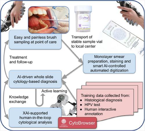
BACKGROUND Oral squamous cell carcinoma (OSCC) is a widespread disease with only 50%-60% 5-year survival. Individuals with potentially malignant precursor lesions are at high risk. METHODS Survival could be increased by effective, affordable, and simple screening methods, along with a shift from incisional tissue biopsies to non-invasive brush biopsies for cytology diagnosis, which are easy to perform in primary care. Along with the explainable, fast, and objective artificial intelligence characterisation of cells through deep learning, an easy-to-use, rapid, and cost-effective methodology for finding high-risk lesions is achievable. The collection of cytology samples offers the further opportunity of explorative genomic analysis. RESULTS Our prospective multicentre study of patients with leukoplakia yields a vast number of oral keratinocytes. In addition to cytopathological analysis, whole-slide imaging and the training of deep neural networks, samples are analysed according to a single-cell RNA sequencing protocol, enabling mapping of the entire keratinocyte transcriptome. Mapping the changes in the genetic profile, based on mRNA expression, facilitates the identification of biomarkers that predict cancer transformation. CONCLUSION This position paper highlights non-invasive methods for identifying patients with oral mucosal lesions at risk of malignant transformation. Reliable non-invasive methods for screening at-risk individuals bring the early diagnosis of OSCC within reach. The use of biomarkers to decide on a targeted therapy is most likely to improve the outcome. With the large-scale collection of samples following patients over time, combined with genomic analysis and modern machine-learning-based approaches for finding patterns in data, this path holds great promise.

口腔鳞状细胞癌预防和诊断的范式转变。
背景:口腔鳞状细胞癌是一种广泛存在的疾病,5年生存率仅为50%-60%。具有潜在恶性前驱病变的个体风险较高。方法:通过有效、负担得起和简单的筛查方法,以及从切口组织活检转向无创刷活检进行细胞学诊断,可以提高生存率,这在初级保健中很容易进行。除了通过深度学习对细胞进行可解释、快速、客观的人工智能表征外,还可以实现一种易于使用、快速、成本效益高的方法来发现高风险病变。细胞学样本的收集为探索性基因组分析提供了进一步的机会。结果:我们对白斑患者的前瞻性多中心研究产生了大量的口腔角质形成细胞。除了细胞病理学分析、全玻片成像和深度神经网络的训练外,还根据单细胞RNA测序方案对样本进行分析,从而能够绘制整个角质形成细胞转录组的图谱。基于mRNA表达绘制基因图谱的变化有助于识别预测癌症转化的生物标志物。结论:这篇立场论文强调了识别口腔粘膜病变有恶变风险的患者的非侵入性方法。筛查高危人群的可靠非侵入性方法使OSCC的早期诊断触手可及。使用生物标志物来决定靶向治疗最有可能改善结果。随着时间的推移,对患者进行大规模的样本采集,再加上基因组分析和基于现代机器学习的方法来寻找数据模式,这条道路前景广阔。
本文章由计算机程序翻译,如有差异,请以英文原文为准。
求助全文
约1分钟内获得全文
求助全文
来源期刊
CiteScore
5.90
自引率
6.10%
发文量
121
审稿时长
4-8 weeks
期刊介绍:
The aim of the Journal of Oral Pathology & Medicine is to publish manuscripts of high scientific quality representing original clinical, diagnostic or experimental work in oral pathology and oral medicine. Papers advancing the science or practice of these disciplines will be welcomed, especially those which bring new knowledge and observations from the application of techniques within the spheres of light and electron microscopy, tissue and organ culture, immunology, histochemistry and immunocytochemistry, microbiology, genetics and biochemistry.

 求助内容:
求助内容: 应助结果提醒方式:


