Ji-Hye Yea, Mario Gomez-Salazar, Sharon Onggo, Zhao Li, Neelima Thottappillil, Masnsen Cherief, Stefano Negri, Xin Xing, Qizhi Qin, Robert Joel Tower, Chen-Ming Fan, Benjamin Levi, Aaron W James
{"title":"Tppp3<sup>+</sup> synovial/tendon sheath progenitor cells contribute to heterotopic bone after trauma.","authors":"Ji-Hye Yea, Mario Gomez-Salazar, Sharon Onggo, Zhao Li, Neelima Thottappillil, Masnsen Cherief, Stefano Negri, Xin Xing, Qizhi Qin, Robert Joel Tower, Chen-Ming Fan, Benjamin Levi, Aaron W James","doi":"10.1038/s41413-023-00272-x","DOIUrl":null,"url":null,"abstract":"<p><p>Heterotopic ossification (HO) is a pathological process resulting in aberrant bone formation and often involves synovial lined tissues. During this process, mesenchymal progenitor cells undergo endochondral ossification. Nonetheless, the specific cell phenotypes and mechanisms driving this process are not well understood, in part due to the high degree of heterogeneity of the progenitor cells involved. Here, using a combination of lineage tracing and single-cell RNA sequencing (scRNA-seq), we investigated the extent to which synovial/tendon sheath progenitor cells contribute to heterotopic bone formation. For this purpose, Tppp3 (tubulin polymerization-promoting protein family member 3)-inducible reporter mice were used in combination with either Scx (Scleraxis) or Pdgfra (platelet derived growth factor receptor alpha) reporter mice. Both tendon injury- and arthroplasty-induced mouse experimental HO models were utilized. ScRNA-seq of tendon-associated traumatic HO suggested that Tppp3 is an early progenitor cell marker for either tendon or osteochondral cells. Upon HO induction, Tppp3 reporter<sup>+</sup> cells expanded in number and partially contributed to cartilage and bone formation in either tendon- or joint-associated HO. In double reporter animals, both Pdgfra<sup>+</sup>Tppp3<sup>+</sup> and Pdgfra<sup>+</sup>Tppp3<sup>-</sup> progenitor cells gave rise to HO-associated cartilage. Finally, analysis of human samples showed a substantial population of TPPP3-expressing cells overlapping with osteogenic markers in areas of heterotopic bone. Overall, these data demonstrate that synovial/tendon sheath progenitor cells undergo aberrant osteochondral differentiation and contribute to HO after trauma.</p>","PeriodicalId":9134,"journal":{"name":"Bone Research","volume":"11 1","pages":"39"},"PeriodicalIF":14.3000,"publicationDate":"2023-07-21","publicationTypes":"Journal Article","fieldsOfStudy":null,"isOpenAccess":false,"openAccessPdf":"https://www.ncbi.nlm.nih.gov/pmc/articles/PMC10361999/pdf/","citationCount":"0","resultStr":null,"platform":"Semanticscholar","paperid":null,"PeriodicalName":"Bone Research","FirstCategoryId":"3","ListUrlMain":"https://doi.org/10.1038/s41413-023-00272-x","RegionNum":1,"RegionCategory":"医学","ArticlePicture":[],"TitleCN":null,"AbstractTextCN":null,"PMCID":null,"EPubDate":"","PubModel":"","JCR":"Q1","JCRName":"CELL & TISSUE ENGINEERING","Score":null,"Total":0}
引用次数: 0
Abstract
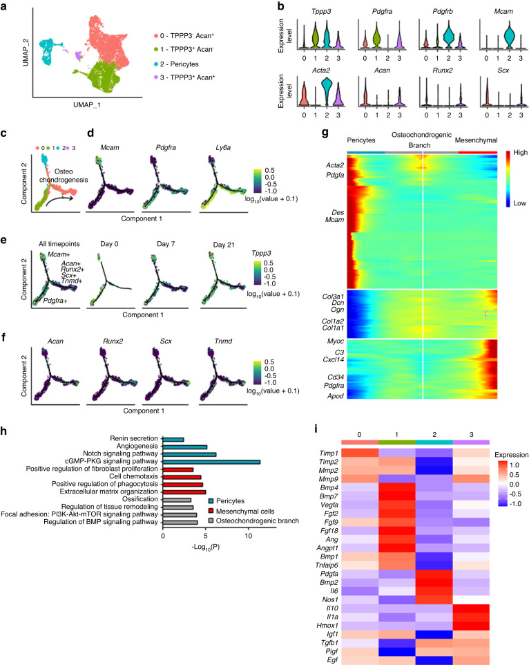
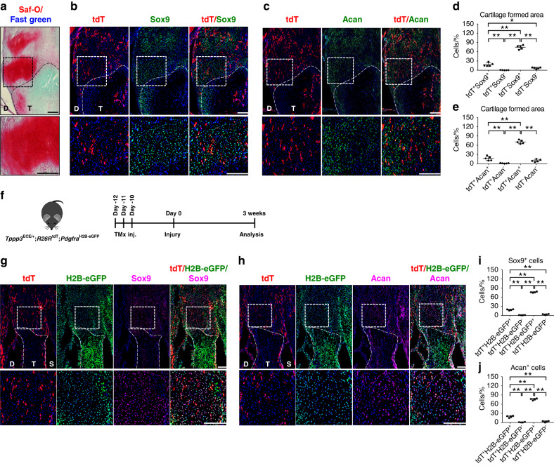
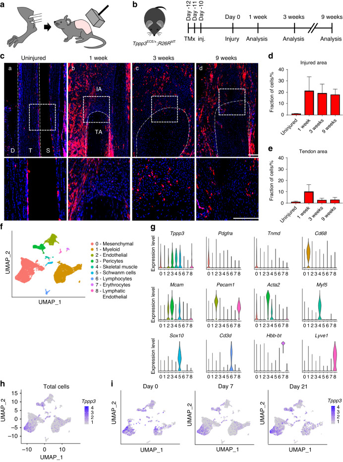
Heterotopic ossification (HO) is a pathological process resulting in aberrant bone formation and often involves synovial lined tissues. During this process, mesenchymal progenitor cells undergo endochondral ossification. Nonetheless, the specific cell phenotypes and mechanisms driving this process are not well understood, in part due to the high degree of heterogeneity of the progenitor cells involved. Here, using a combination of lineage tracing and single-cell RNA sequencing (scRNA-seq), we investigated the extent to which synovial/tendon sheath progenitor cells contribute to heterotopic bone formation. For this purpose, Tppp3 (tubulin polymerization-promoting protein family member 3)-inducible reporter mice were used in combination with either Scx (Scleraxis) or Pdgfra (platelet derived growth factor receptor alpha) reporter mice. Both tendon injury- and arthroplasty-induced mouse experimental HO models were utilized. ScRNA-seq of tendon-associated traumatic HO suggested that Tppp3 is an early progenitor cell marker for either tendon or osteochondral cells. Upon HO induction, Tppp3 reporter+ cells expanded in number and partially contributed to cartilage and bone formation in either tendon- or joint-associated HO. In double reporter animals, both Pdgfra+Tppp3+ and Pdgfra+Tppp3- progenitor cells gave rise to HO-associated cartilage. Finally, analysis of human samples showed a substantial population of TPPP3-expressing cells overlapping with osteogenic markers in areas of heterotopic bone. Overall, these data demonstrate that synovial/tendon sheath progenitor cells undergo aberrant osteochondral differentiation and contribute to HO after trauma.
期刊介绍:
Established in 2013, Bone Research is a newly-founded English-language periodical that centers on the basic and clinical facets of bone biology, pathophysiology, and regeneration. It is dedicated to championing key findings emerging from both basic investigations and clinical research concerning bone-related topics. The journal's objective is to globally disseminate research in bone-related physiology, pathology, diseases, and treatment, contributing to the advancement of knowledge in this field.
 求助内容:
求助内容: 应助结果提醒方式:


