{"title":"Clinical Course and Optical Coherence Tomography of Ocular Angiostrongyliasis: A Case Report.","authors":"Kanin Luangsawang, Veeraphatra Wongsantimeth, Sirinan Treeyawedkul","doi":"10.4103/joco.joco_137_22","DOIUrl":null,"url":null,"abstract":"<p><strong>Purpose: </strong>To report the clinical course and optical coherence tomography (OCT) findings of ocular angiostrongyliasis.</p><p><strong>Methods: </strong>A 36-year-old female with a history of ingesting regular raw freshwater shrimp and other raw food presented with acute unilateral painless visual loss in the right eye. Her right eye's best-corrected visual acuity (BCVA) was 1 ft of the count finger. Fundus examination showed vitritis, generalized retinal pigment epithelial alteration, and a moving roundworm in the vitreous at the 6 o'clock position. Macular OCT of her right eye showed thinning of the retina, loss of the external limiting membrane and ellipsoid zone, subretinal hyper-reflective material clumping, and hyper-reflective foci at the superficial choroidal layer.</p><p><strong>Results: </strong>The patient was administered oral and topical prednisolone. The roundworm, identified as <i>Angiostrongylus cantonensis</i>, was wholly extracted from the vitreous using a 23G sclerotomy port and pars plana vitrectomy. The final BCVA was 1 ft of the count finger.</p><p><strong>Conclusion: </strong>This case report describes an infrequent presentation and illustrates the clinical course and OCT findings of ocular angiostrongyliasis.</p>","PeriodicalId":15423,"journal":{"name":"Journal of Current Ophthalmology","volume":"35 1","pages":"86-89"},"PeriodicalIF":1.2000,"publicationDate":"2023-01-01","publicationTypes":"Journal Article","fieldsOfStudy":null,"isOpenAccess":false,"openAccessPdf":"https://ftp.ncbi.nlm.nih.gov/pub/pmc/oa_pdf/0a/12/JCO-35-86.PMC10481986.pdf","citationCount":"0","resultStr":null,"platform":"Semanticscholar","paperid":null,"PeriodicalName":"Journal of Current Ophthalmology","FirstCategoryId":"1085","ListUrlMain":"https://doi.org/10.4103/joco.joco_137_22","RegionNum":0,"RegionCategory":null,"ArticlePicture":[],"TitleCN":null,"AbstractTextCN":null,"PMCID":null,"EPubDate":"","PubModel":"","JCR":"Q3","JCRName":"OPHTHALMOLOGY","Score":null,"Total":0}
引用次数: 0
Abstract
Purpose: To report the clinical course and optical coherence tomography (OCT) findings of ocular angiostrongyliasis.
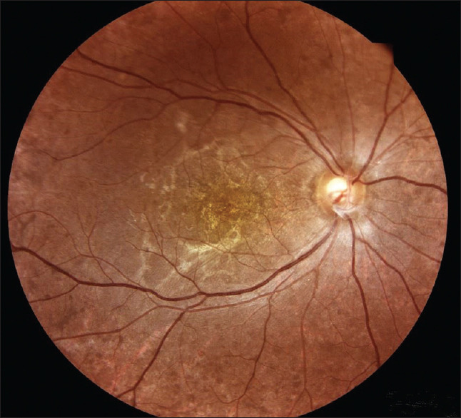
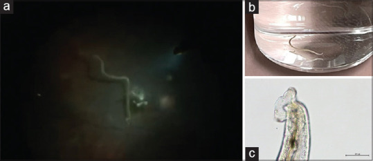
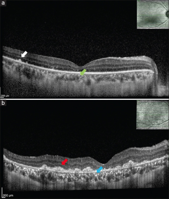
Methods: A 36-year-old female with a history of ingesting regular raw freshwater shrimp and other raw food presented with acute unilateral painless visual loss in the right eye. Her right eye's best-corrected visual acuity (BCVA) was 1 ft of the count finger. Fundus examination showed vitritis, generalized retinal pigment epithelial alteration, and a moving roundworm in the vitreous at the 6 o'clock position. Macular OCT of her right eye showed thinning of the retina, loss of the external limiting membrane and ellipsoid zone, subretinal hyper-reflective material clumping, and hyper-reflective foci at the superficial choroidal layer.
Results: The patient was administered oral and topical prednisolone. The roundworm, identified as Angiostrongylus cantonensis, was wholly extracted from the vitreous using a 23G sclerotomy port and pars plana vitrectomy. The final BCVA was 1 ft of the count finger.
Conclusion: This case report describes an infrequent presentation and illustrates the clinical course and OCT findings of ocular angiostrongyliasis.
期刊介绍:
Peer Review under the responsibility of Iranian Society of Ophthalmology Journal of Current Ophthalmology, the official publication of the Iranian Society of Ophthalmology, is a peer-reviewed, open-access, scientific journal that welcomes high quality original articles related to vision science and all fields of ophthalmology. Journal of Current Ophthalmology is the continuum of Iranian Journal of Ophthalmology published since 1969.

 求助内容:
求助内容: 应助结果提醒方式:


