Mohammad Naser Hashemian, Sadegh Ghafarian, Hamid Riazi-Esfahani, Elias Khalili Pour
{"title":"Evaluation of Choroidal Vascularity Index in Keratoconus Patients: Does Choroidal Vascularity Change in Keratoconus?","authors":"Mohammad Naser Hashemian, Sadegh Ghafarian, Hamid Riazi-Esfahani, Elias Khalili Pour","doi":"10.4103/joco.joco_189_22","DOIUrl":null,"url":null,"abstract":"<p><strong>Purpose: </strong>To investigate the choroidal structure in keratoconic patients with different severity using the choroidal vascularity index (CVI) derived from image binarization on enhanced depth imaging optical coherence tomography scans (EDI-OCT).</p><p><strong>Methods: </strong>Sixty-eight eyes from 34 keratoconus (KCN) patients and 72 eyes from 36 healthy subjects were recruited in this prospective, noninterventional, comparative cross-sectional study. EDI-OCT was employed to measure choroidal parameters, including choroidal thickness (CT), total choroidal area (TCA), luminal area, stromal area, and CVI.</p><p><strong>Results: </strong>Subfoveal CT was 354.6 ± 66.8 μm in the control group and 371 ± 64.5 μm in the KCN group (<i>P</i> = 0.86). There was no significant difference between control and KCN groups in terms of TCA (0.66 ± 0.14 mm<sup>2</sup> vs. 0.7 ± 0.12 mm<sup>2</sup>; <i>P</i> = 0.70), luminal area (0.49 ± 0.10 mm<sup>2</sup> vs. 0.53 ± 0.08 mm<sup>2</sup>; <i>P</i> = 0.67), and stromal area (0.16 ± 0.05 mm<sup>2</sup> vs. 0.17 ± 0.05 mm<sup>2</sup>; <i>P</i> = 0.84). CVI was also comparable in the control group (75.4% ±3.4%) and the KCN group (75.6% ±4.5%; <i>P</i> = 0.43). There was also no significant correlation between other choroidal parameters and KCN severity indices.</p><p><strong>Conclusion: </strong>It seems that CVI as well as other choroidal biomarkers were not significantly different between patients with KCN and healthy subjects.</p>","PeriodicalId":15423,"journal":{"name":"Journal of Current Ophthalmology","volume":"35 1","pages":"36-41"},"PeriodicalIF":1.2000,"publicationDate":"2023-01-01","publicationTypes":"Journal Article","fieldsOfStudy":null,"isOpenAccess":false,"openAccessPdf":"https://ftp.ncbi.nlm.nih.gov/pub/pmc/oa_pdf/2e/3d/JCO-35-36.PMC10481970.pdf","citationCount":"0","resultStr":null,"platform":"Semanticscholar","paperid":null,"PeriodicalName":"Journal of Current Ophthalmology","FirstCategoryId":"1085","ListUrlMain":"https://doi.org/10.4103/joco.joco_189_22","RegionNum":0,"RegionCategory":null,"ArticlePicture":[],"TitleCN":null,"AbstractTextCN":null,"PMCID":null,"EPubDate":"","PubModel":"","JCR":"Q3","JCRName":"OPHTHALMOLOGY","Score":null,"Total":0}
引用次数: 0
Abstract
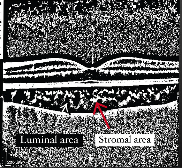
Purpose: To investigate the choroidal structure in keratoconic patients with different severity using the choroidal vascularity index (CVI) derived from image binarization on enhanced depth imaging optical coherence tomography scans (EDI-OCT).
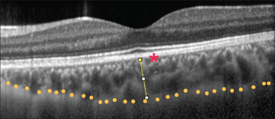
Methods: Sixty-eight eyes from 34 keratoconus (KCN) patients and 72 eyes from 36 healthy subjects were recruited in this prospective, noninterventional, comparative cross-sectional study. EDI-OCT was employed to measure choroidal parameters, including choroidal thickness (CT), total choroidal area (TCA), luminal area, stromal area, and CVI.
Results: Subfoveal CT was 354.6 ± 66.8 μm in the control group and 371 ± 64.5 μm in the KCN group (P = 0.86). There was no significant difference between control and KCN groups in terms of TCA (0.66 ± 0.14 mm2 vs. 0.7 ± 0.12 mm2; P = 0.70), luminal area (0.49 ± 0.10 mm2 vs. 0.53 ± 0.08 mm2; P = 0.67), and stromal area (0.16 ± 0.05 mm2 vs. 0.17 ± 0.05 mm2; P = 0.84). CVI was also comparable in the control group (75.4% ±3.4%) and the KCN group (75.6% ±4.5%; P = 0.43). There was also no significant correlation between other choroidal parameters and KCN severity indices.
Conclusion: It seems that CVI as well as other choroidal biomarkers were not significantly different between patients with KCN and healthy subjects.
期刊介绍:
Peer Review under the responsibility of Iranian Society of Ophthalmology Journal of Current Ophthalmology, the official publication of the Iranian Society of Ophthalmology, is a peer-reviewed, open-access, scientific journal that welcomes high quality original articles related to vision science and all fields of ophthalmology. Journal of Current Ophthalmology is the continuum of Iranian Journal of Ophthalmology published since 1969.

 求助内容:
求助内容: 应助结果提醒方式:


