Three-dimensional evaluation of alveolar bone and pharyngeal airway dimensions after mandibular dentition distalization in patients with Class III malocclusion: a retrospective study.
{"title":"Three-dimensional evaluation of alveolar bone and pharyngeal airway dimensions after mandibular dentition distalization in patients with Class III malocclusion: a retrospective study.","authors":"Zhijie Zhou, Liangyan Sun, Fan Zhang, Yan Xu","doi":"10.1186/s13005-023-00382-1","DOIUrl":null,"url":null,"abstract":"<p><strong>Background: </strong>To three-dimensionally evaluate changes of the alveolar bone around the mandibular anterior teeth and pharyngeal airway dimensions in adults with Class III malocclusion before and after orthodontic treatment of mandibular dentition distalization.</p><p><strong>Methods: </strong>In this retrospective study, cone-beam computed tomography (CBCT) scans of 20 patients with Class III malocclusion who underwent mandibular dentition distalization were obtained both before and after treatment. Three-dimensional changes of the thickness and vertical marginal bone levels around mandibular incisors and canines were assessed and compared. And airway volumes of the palato-, glosso-, laryngopharynx and the minimum axial area were measured and compared before and after treatment.</p><p><strong>Results: </strong>A significant decrease of lingual bone thickness of mandibular incisors, partial labial and lingual bone thickness of canines were observed (P < 0.05). The reduction in root length of incisors and canines, labial and lingual vertical marginal bone levels were significant after orthodontic treatment. No significant correlations between mandibular dentition distalization and pharyngeal airway dimensions were observed.</p><p><strong>Conclusions: </strong>Mandibular dentition distalization could result in the loss of alveolar bone around anterior teeth in Class III malocclusion, especially for the cervical marginal bone. Pharyngeal airway dimensions were not affected to a high extent after distalization.</p><p><strong>Trial registration: </strong>Retrospctively registered.</p>","PeriodicalId":12994,"journal":{"name":"Head & Face Medicine","volume":null,"pages":null},"PeriodicalIF":2.4000,"publicationDate":"2023-08-30","publicationTypes":"Journal Article","fieldsOfStudy":null,"isOpenAccess":false,"openAccessPdf":"https://www.ncbi.nlm.nih.gov/pmc/articles/PMC10466725/pdf/","citationCount":"0","resultStr":null,"platform":"Semanticscholar","paperid":null,"PeriodicalName":"Head & Face Medicine","FirstCategoryId":"3","ListUrlMain":"https://doi.org/10.1186/s13005-023-00382-1","RegionNum":2,"RegionCategory":"医学","ArticlePicture":[],"TitleCN":null,"AbstractTextCN":null,"PMCID":null,"EPubDate":"","PubModel":"","JCR":"Q2","JCRName":"DENTISTRY, ORAL SURGERY & MEDICINE","Score":null,"Total":0}
引用次数: 0
Abstract
Background: To three-dimensionally evaluate changes of the alveolar bone around the mandibular anterior teeth and pharyngeal airway dimensions in adults with Class III malocclusion before and after orthodontic treatment of mandibular dentition distalization.
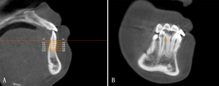
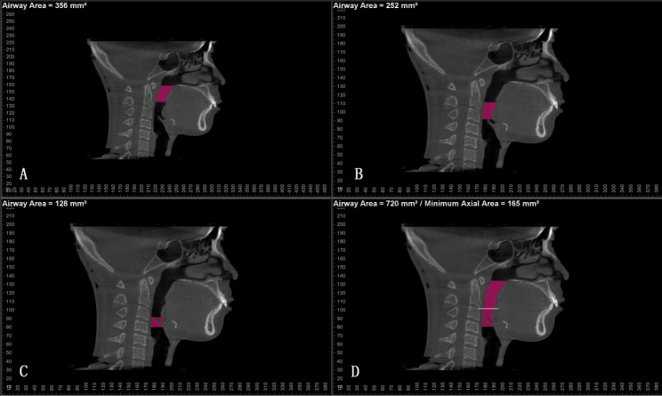
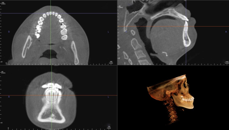
Methods: In this retrospective study, cone-beam computed tomography (CBCT) scans of 20 patients with Class III malocclusion who underwent mandibular dentition distalization were obtained both before and after treatment. Three-dimensional changes of the thickness and vertical marginal bone levels around mandibular incisors and canines were assessed and compared. And airway volumes of the palato-, glosso-, laryngopharynx and the minimum axial area were measured and compared before and after treatment.
Results: A significant decrease of lingual bone thickness of mandibular incisors, partial labial and lingual bone thickness of canines were observed (P < 0.05). The reduction in root length of incisors and canines, labial and lingual vertical marginal bone levels were significant after orthodontic treatment. No significant correlations between mandibular dentition distalization and pharyngeal airway dimensions were observed.
Conclusions: Mandibular dentition distalization could result in the loss of alveolar bone around anterior teeth in Class III malocclusion, especially for the cervical marginal bone. Pharyngeal airway dimensions were not affected to a high extent after distalization.
期刊介绍:
Head & Face Medicine is a multidisciplinary open access journal that publishes basic and clinical research concerning all aspects of cranial, facial and oral conditions.
The journal covers all aspects of cranial, facial and oral diseases and their management. It has been designed as a multidisciplinary journal for clinicians and researchers involved in the diagnostic and therapeutic aspects of diseases which affect the human head and face. The journal is wide-ranging, covering the development, aetiology, epidemiology and therapy of head and face diseases to the basic science that underlies these diseases. Management of head and face diseases includes all aspects of surgical and non-surgical treatments including psychopharmacological therapies.
 求助内容:
求助内容: 应助结果提醒方式:


