Mizuho Kittaka, Tetsuya Yoshimoto, Marcus E Levitan, Rina Urata, Roy B Choi, Yayoi Teno, Yixia Xie, Yukiko Kitase, Matthew Prideaux, Sarah L Dallas, Alexander G Robling, Yasuyoshi Ueki
下载PDF
{"title":"Osteocyte RANKL Drives Bone Resorption in Mouse Ligature-Induced Periodontitis","authors":"Mizuho Kittaka, Tetsuya Yoshimoto, Marcus E Levitan, Rina Urata, Roy B Choi, Yayoi Teno, Yixia Xie, Yukiko Kitase, Matthew Prideaux, Sarah L Dallas, Alexander G Robling, Yasuyoshi Ueki","doi":"10.1002/jbmr.4897","DOIUrl":null,"url":null,"abstract":"<p>Mouse ligature-induced periodontitis (LIP) has been used to study bone loss in periodontitis. However, the role of osteocytes in LIP remains unclear. Furthermore, there is no consensus on the choice of alveolar bone parameters and time points to evaluate LIP. Here, we investigated the dynamics of changes in osteoclastogenesis and bone volume (BV) loss in LIP over 14 days. Time-course analysis revealed that osteoclast induction peaked on days 3 and 5, followed by the peak of BV loss on day 7. Notably, BV was restored by day 14. The bone formation phase after the bone resorption phase was suggested to be responsible for the recovery of bone loss. Electron microscopy identified bacteria in the osteocyte lacunar space beyond the periodontal ligament (PDL) tissue. We investigated how osteocytes affect bone resorption of LIP and found that mice lacking receptor activator of NF-κB ligand (RANKL), predominantly in osteocytes, protected against bone loss in LIP, whereas recombination activating 1 (RAG1)-deficient mice failed to resist it. These results indicate that T/B cells are dispensable for osteoclast induction in LIP and that RANKL from osteocytes and mature osteoblasts regulates bone resorption by LIP. Remarkably, mice lacking the myeloid differentiation primary response gene 88 (MYD88) did not show protection against LIP-induced bone loss. Instead, osteocytic cells expressed nucleotide-binding oligomerization domain containing 1 (NOD1), and primary osteocytes induced significantly higher <i>Rankl</i> than primary osteoblasts when stimulated with a NOD1 agonist. Taken together, LIP induced both bone resorption and bone formation in a stage-dependent manner, suggesting that the selection of time points is critical for quantifying bone loss in mouse LIP. Pathogenetically, the current study suggests that bacterial activation of osteocytes via NOD1 is involved in the mechanism of osteoclastogenesis in LIP. The NOD1-RANKL axis in osteocytes may be a therapeutic target for bone resorption in periodontitis. © 2023 The Authors. <i>Journal of Bone and Mineral Research</i> published by Wiley Periodicals LLC on behalf of American Society for Bone and Mineral Research (ASBMR).</p>","PeriodicalId":185,"journal":{"name":"Journal of Bone and Mineral Research","volume":"38 10","pages":"1521-1540"},"PeriodicalIF":5.1000,"publicationDate":"2023-08-08","publicationTypes":"Journal Article","fieldsOfStudy":null,"isOpenAccess":false,"openAccessPdf":"https://onlinelibrary.wiley.com/doi/epdf/10.1002/jbmr.4897","citationCount":"0","resultStr":null,"platform":"Semanticscholar","paperid":null,"PeriodicalName":"Journal of Bone and Mineral Research","FirstCategoryId":"3","ListUrlMain":"https://onlinelibrary.wiley.com/doi/10.1002/jbmr.4897","RegionNum":1,"RegionCategory":"医学","ArticlePicture":[],"TitleCN":null,"AbstractTextCN":null,"PMCID":null,"EPubDate":"","PubModel":"","JCR":"Q1","JCRName":"ENDOCRINOLOGY & METABOLISM","Score":null,"Total":0}
引用次数: 0
引用
批量引用
Abstract
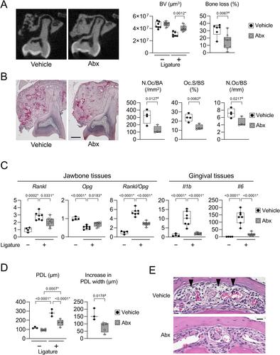
Mouse ligature-induced periodontitis (LIP) has been used to study bone loss in periodontitis. However, the role of osteocytes in LIP remains unclear. Furthermore, there is no consensus on the choice of alveolar bone parameters and time points to evaluate LIP. Here, we investigated the dynamics of changes in osteoclastogenesis and bone volume (BV) loss in LIP over 14 days. Time-course analysis revealed that osteoclast induction peaked on days 3 and 5, followed by the peak of BV loss on day 7. Notably, BV was restored by day 14. The bone formation phase after the bone resorption phase was suggested to be responsible for the recovery of bone loss. Electron microscopy identified bacteria in the osteocyte lacunar space beyond the periodontal ligament (PDL) tissue. We investigated how osteocytes affect bone resorption of LIP and found that mice lacking receptor activator of NF-κB ligand (RANKL), predominantly in osteocytes, protected against bone loss in LIP, whereas recombination activating 1 (RAG1)-deficient mice failed to resist it. These results indicate that T/B cells are dispensable for osteoclast induction in LIP and that RANKL from osteocytes and mature osteoblasts regulates bone resorption by LIP. Remarkably, mice lacking the myeloid differentiation primary response gene 88 (MYD88) did not show protection against LIP-induced bone loss. Instead, osteocytic cells expressed nucleotide-binding oligomerization domain containing 1 (NOD1), and primary osteocytes induced significantly higher Rankl than primary osteoblasts when stimulated with a NOD1 agonist. Taken together, LIP induced both bone resorption and bone formation in a stage-dependent manner, suggesting that the selection of time points is critical for quantifying bone loss in mouse LIP. Pathogenetically, the current study suggests that bacterial activation of osteocytes via NOD1 is involved in the mechanism of osteoclastogenesis in LIP. The NOD1-RANKL axis in osteocytes may be a therapeutic target for bone resorption in periodontitis. © 2023 The Authors. Journal of Bone and Mineral Research published by Wiley Periodicals LLC on behalf of American Society for Bone and Mineral Research (ASBMR).
骨细胞RANKL在小鼠连字诱导的牙周炎中驱动骨吸收
小鼠结扎诱导牙周炎(LIP)已被用于研究牙周炎中的骨丢失。然而,骨细胞在LIP中的作用尚不清楚。此外,对于评估LIP的牙槽骨参数和时间点的选择,还没有达成共识。在这里,我们研究了14岁以上LIP破骨细胞生成和骨体积(BV)损失的动态变化 天。时间进程分析显示,破骨细胞诱导在第3天和第5天达到峰值,随后BV损失在第7天达到峰值。值得注意的是,BV在第14天恢复。骨吸收阶段之后的骨形成阶段被认为是骨丢失恢复的原因。电子显微镜在牙周膜(PDL)组织外的骨细胞腔隙中发现了细菌。我们研究了骨细胞如何影响LIP的骨吸收,发现缺乏NF-κB配体受体激活剂(RANKL)的小鼠(主要在骨细胞中)可以防止LIP中的骨丢失,而重组激活1(RAG1)缺乏的小鼠无法抵抗。这些结果表明,T/B细胞对于LIP中的破骨细胞诱导是可有可无的,并且来自骨细胞和成熟成骨细胞的RANKL通过LIP调节骨吸收。值得注意的是,缺乏骨髓分化初级反应基因88(MYD88)的小鼠对LIP诱导的骨丢失没有表现出保护作用。相反,骨细胞表达含有核苷酸结合寡聚化结构域1(NOD1),并且当用NOD1激动剂刺激时,原代骨细胞诱导的Rankl显著高于原代成骨细胞。总之,LIP以阶段依赖的方式诱导骨吸收和骨形成,这表明时间点的选择对于量化小鼠LIP中的骨损失至关重要。从发病机制上讲,目前的研究表明,细菌通过NOD1激活骨细胞参与了LIP破骨细胞生成的机制。骨细胞中的NOD1-RANKL轴可能是牙周炎中骨吸收的治疗靶点。©2023作者。由Wiley Periodicals LLC代表美国骨与矿物研究学会(ASBMR)出版的《骨与矿产研究杂志》。
本文章由计算机程序翻译,如有差异,请以英文原文为准。


 求助内容:
求助内容: 应助结果提醒方式:


